Department of Diagnostic and Interventional Radiology, University of Heidelberg, Heidelberg, Germany.
Translational Lung Research Center, Member of the German Center for Lung Research, University of Heidelberg, Heidelberg, Germany.
PLoS One. 2020 Sep 17;15(9):e0238798. doi: 10.1371/journal.pone.0238798. eCollection 2020.
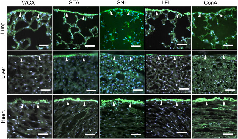
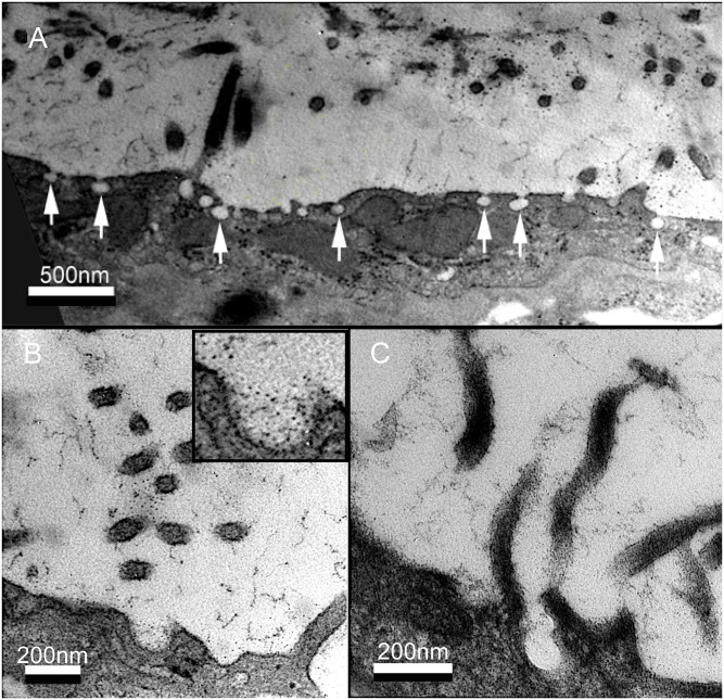
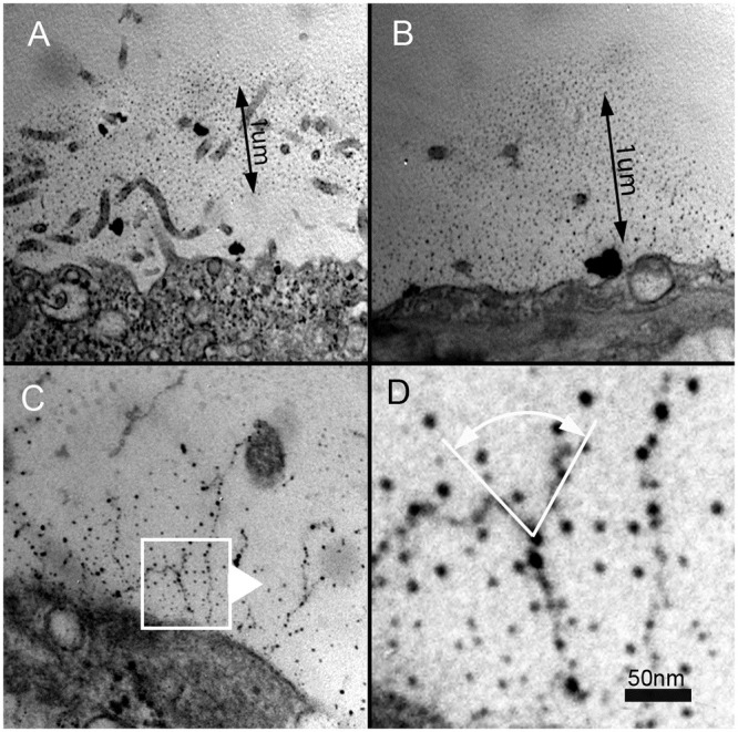
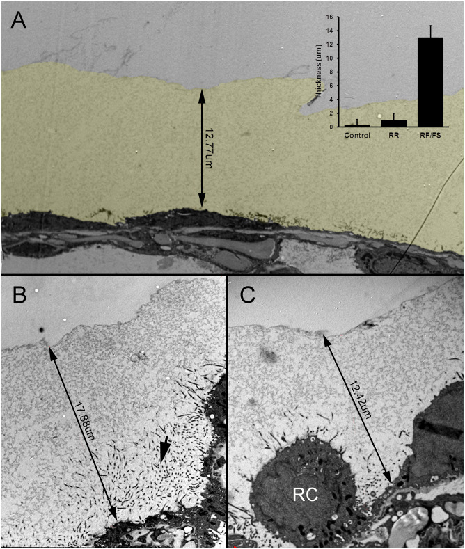
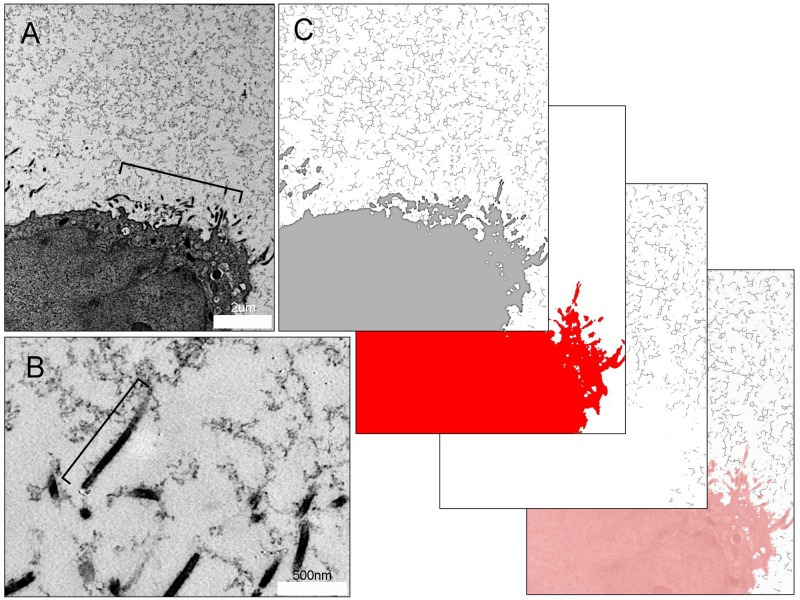
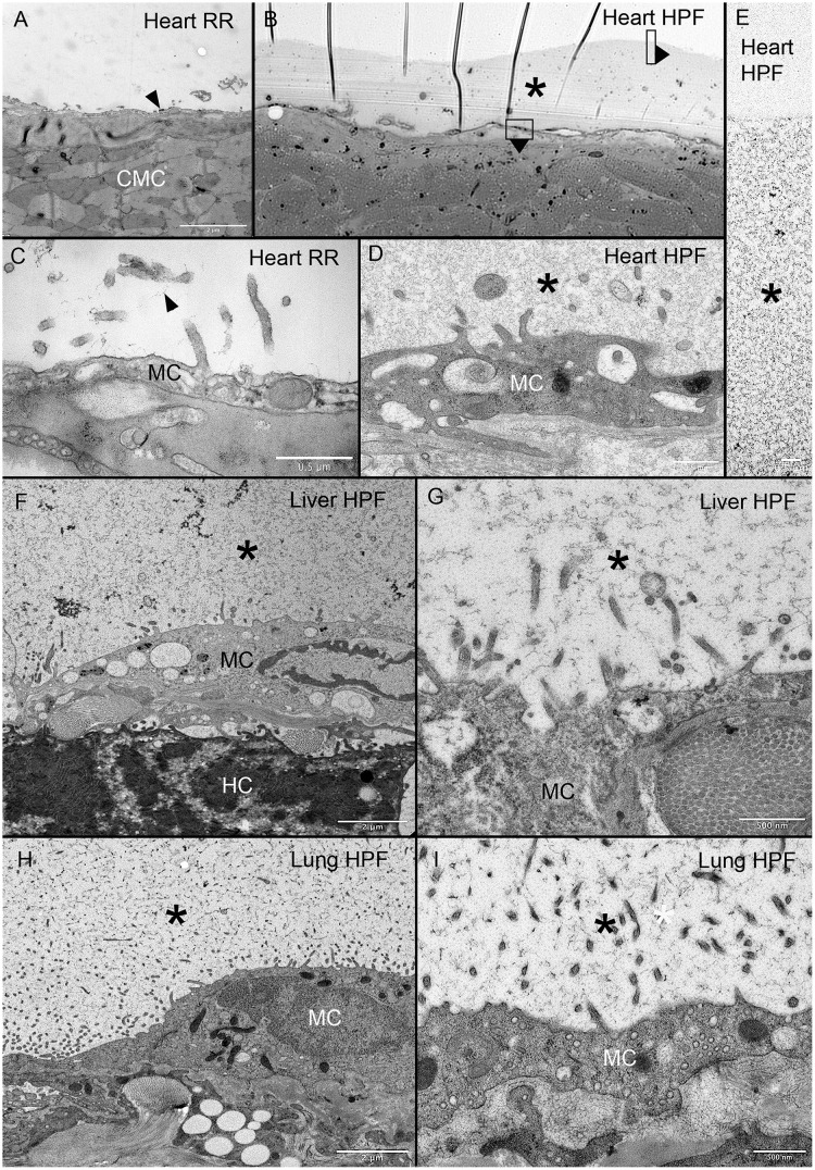
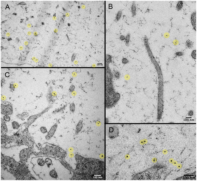
The mesothelium is a dynamic and specialized tissue layer that covers the somatic cavities (pleural, peritoneal, and pericardial) as well as the surface of the visceral organs such as the lung, heart, liver, bowel and tunica vaginalis testis. The potential therapeutic manipulation of visceral organs has been complicated by the carbohydrate surface layer-here, called the mesopolysaccharide (MPS)-that coats the outer layer of the mesothelium. The traditional understanding of MPS structure has relied upon fixation techniques known to degrade carbohydrates. The recent development of carbohydrate-preserving fixation for high resolution imaging techniques has provided an opportunity to re-examine the structure of both the MPS and the visceral mesothelium. In this report, we used high pressure freezing (HPF) as well as serial section transmission electron microscopy to redefine the structure of the MPS expressed on the murine lung, heart and liver surface. Tissue preserved by HPF and examined by transmission electron microscopy demonstrated a pleural MPS layer 13.01±1.1 um deep-a 100-fold increase in depth compared to previously reported data obtained with conventional fixation techniques. At the base of the MPS were microvilli 1.1±0.35 um long and 42±5 nm in diameter. Morphological evidence suggested that the MPS was anchored to the mesothelium by microvilli. In addition, membrane pits 97±17 nm in diameter were observed in the apical mesothelial membrane. The spatial proximity and surface density (29±4.5%) of the pits suggested an active process linked to the structural maintenance of the MPS. The striking magnitude and complex structure of the MPS indicates that it is an important consideration in studies of the visceral mesothelium.
间皮是一种动态且特化的组织层,覆盖体腔(胸膜、腹膜和心包)以及内脏器官的表面,如肺、心脏、肝脏、肠道和睾丸鞘膜。由于覆盖间皮外层的碳水化合物表面层,即间多糖(MPS),对内脏器官的潜在治疗操作变得复杂。传统上对 MPS 结构的理解依赖于已知会降解碳水化合物的固定技术。用于高分辨率成像技术的碳水化合物保存固定技术的最新发展提供了重新检查 MPS 和内脏间皮结构的机会。在本报告中,我们使用高压冷冻(HPF)以及连续切片透射电子显微镜来重新定义在小鼠肺、心脏和肝脏表面表达的 MPS 的结构。通过 HPF 保存并通过透射电子显微镜检查的组织显示胸膜 MPS 层深 13.01±1.1um-与传统固定技术获得的先前报告数据相比,深度增加了 100 倍。在 MPS 的底部是长 1.1±0.35um 且直径为 42±5nm 的微绒毛。形态学证据表明,MPS 通过微绒毛锚定到间皮上。此外,在顶膜上皮细胞的顶端膜上观察到直径为 97±17nm 的膜陷窝。陷窝的空间接近度和表面密度(29±4.5%)表明这是与 MPS 结构维持相关的一个活跃过程。MPS 的显著幅度和复杂结构表明,在研究内脏间皮时,这是一个重要的考虑因素。