Benjamin Christopher F, Walshaw Patricia D, Hale Kayleigh, Gaillard William D, Baxter Leslie C, Berl Madison M, Polczynska Monika, Noble Stephanie, Alkawadri Rafeed, Hirsch Lawrence J, Constable R Todd, Bookheimer Susan Y
Department of Neurology, Comprehensive Epilepsy Center, Yale School of Medicine, New Haven, Connecticut.
Department of Neurosurgery, Yale School of Medicine, New Haven, Connecticut.
Hum Brain Mapp. 2017 Aug;38(8):4239-4255. doi: 10.1002/hbm.23661. Epub 2017 May 23.
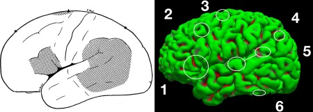
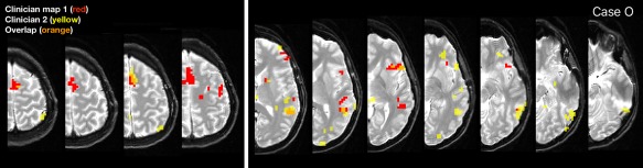
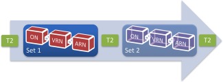
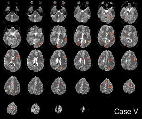
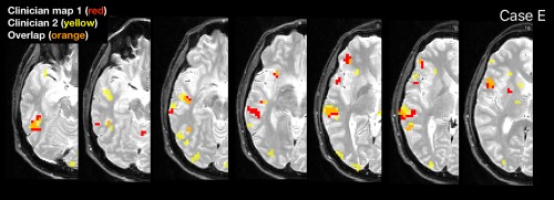
Language mapping is a key goal in neurosurgical planning. fMRI mapping typically proceeds with a focus on Broca's and Wernicke's areas, although multiple other language-critical areas are now well-known. We evaluated whether clinicians could use a novel approach, including clinician-driven individualized thresholding, to reliably identify six language regions, including Broca's Area, Wernicke's Area (inferior, superior), Exner's Area, Supplementary Speech Area, Angular Gyrus, and Basal Temporal Language Area. We studied 22 epilepsy and tumor patients who received Wada and fMRI (age 36.4[12.5]; Wada language left/right/mixed in 18/3/1). fMRI tasks (two × three tasks) were analyzed by two clinical neuropsychologists who flexibly thresholded and combined these to identify the six regions. The resulting maps were compared to fixed threshold maps. Clinicians generated maps that overlapped significantly, and were highly consistent, when at least one task came from the same set. Cases diverged when clinicians prioritized different language regions or addressed noise differently. Language laterality closely mirrored Wada data (85% accuracy). Activation consistent with all six language regions was consistently identified. In blind review, three external, independent clinicians rated the individualized fMRI language maps as superior to fixed threshold maps; identified the majority of regions significantly more frequently; and judged language laterality to mirror Wada lateralization more often. These data provide initial validation of a novel, clinician-based approach to localizing language cortex. They also demonstrate clinical fMRI is superior when analyzed by an experienced clinician and that when fMRI data is of low quality judgments of laterality are unreliable and should be withheld. Hum Brain Mapp 38:4239-4255, 2017. © 2017 Wiley Periodicals, Inc.
语言映射是神经外科手术规划中的一个关键目标。功能磁共振成像(fMRI)映射通常着重于布洛卡区和韦尼克区,不过现在人们已经熟知多个其他对语言至关重要的区域。我们评估了临床医生是否能够使用一种新颖的方法,包括临床医生驱动的个体化阈值设定,来可靠地识别六个语言区域,即布洛卡区、韦尼克区(包括上韦尼克区和下韦尼克区)、埃克斯纳区、补充言语区、角回和颞叶基底语言区。我们研究了22名接受了瓦达测试和功能磁共振成像检查的癫痫和肿瘤患者(年龄36.4[12.5];瓦达测试显示语言功能位于左侧/右侧/双侧混合的患者分别有18例/3例/1例)。两名临床神经心理学家对功能磁共振成像任务(两个×三个任务)进行分析,他们灵活地设定阈值并将其组合起来以识别这六个区域。将生成的图谱与固定阈值图谱进行比较。当至少有一个任务来自同一组时,临床医生生成图谱的重叠度显著,且高度一致。当临床医生优先考虑不同的语言区域或对噪声的处理方式不同时,情况会出现差异。语言偏侧性与瓦达测试数据高度吻合(准确率85%)。始终能识别出与所有六个语言区域一致的激活情况。在盲法评审中,三名外部独立临床医生将个体化的功能磁共振成像语言图谱评为优于固定阈值图谱;更频繁地显著识别出大多数区域;并且判断语言偏侧性更常与瓦达测试的偏侧化情况相符。这些数据为一种基于临床医生的新颖语言皮层定位方法提供了初步验证。它们还表明,经经验丰富的临床医生分析时,临床功能磁共振成像更具优势,并且当功能磁共振成像数据质量较低时,对偏侧性的判断不可靠,应不予采用。《人类大脑图谱》38:4239 - 4255,2017年。© 2017威利期刊公司