Miri Maliheh, Amini Zahra, Rabbani Hossein, Kafieh Raheleh
Electrical Engineering Department, Faculty of Engineering, Higher Educational Complex of Saravan, Saravan, Iran.
Student Research Committee, School of Advanced Technologies in Medicine, Isfahan University of Medical Sciences, Isfahan, Iran.
J Med Signals Sens. 2017 Apr-Jun;7(2):59-70.
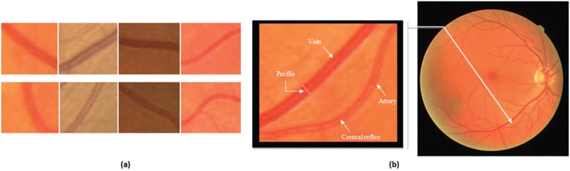
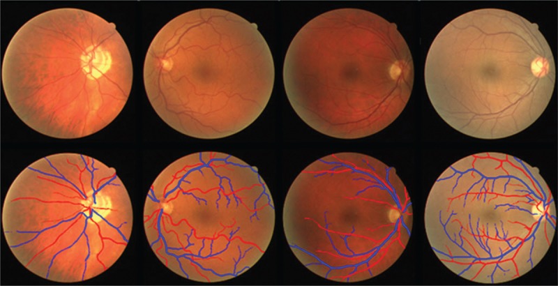
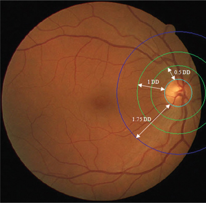
Nowadays, it is obvious that there is a relationship between changes in the retinal vessel structure and diseases such as diabetic, hypertension, stroke, and the other cardiovascular diseases in adults as well as retinopathy of prematurity in infants. Retinal fundus images provide non-invasive visualization of the retinal vessel structure. Applying image processing techniques in the study of digital color fundus photographs and analyzing their vasculature is a reliable approach for early diagnosis of the aforementioned diseases. Reduction in the arteriolar-venular ratio of retina is one of the primary signs of hypertension, diabetic, and cardiovascular diseases which can be calculated by analyzing the fundus images. To achieve a precise measuring of this parameter and meaningful diagnostic results, accurate classification of arteries and veins is necessary. Classification of vessels in fundus images faces with some challenges that make it difficult. In this paper, a comprehensive study of the proposed methods for classification of arteries and veins in fundus images is presented. Considering that these methods are evaluated on different datasets and use different evaluation criteria, it is not possible to conduct a fair comparison of their performance. Therefore, we evaluate the classification methods from modeling perspective. This analysis reveals that most of the proposed approaches have focused on statistics, and geometric models in spatial domain and transform domain models have received less attention. This could suggest the possibility of using transform models, especially data adaptive ones, for modeling of the fundus images in future classification approaches.
如今,视网膜血管结构的变化与糖尿病、高血压、中风等疾病以及成人的其他心血管疾病和婴儿的早产儿视网膜病变之间存在关联,这一点已十分明显。眼底图像提供了视网膜血管结构的非侵入性可视化。在数字彩色眼底照片研究中应用图像处理技术并分析其脉管系统是早期诊断上述疾病的可靠方法。视网膜动静脉比降低是高血压、糖尿病和心血管疾病的主要体征之一,可通过分析眼底图像来计算。为了精确测量该参数并获得有意义的诊断结果,动脉和静脉的准确分类是必要的。眼底图像中的血管分类面临一些挑战,这使其变得困难。本文对眼底图像中动脉和静脉分类的现有方法进行了全面研究。鉴于这些方法在不同数据集上进行评估且使用不同的评估标准,无法对它们的性能进行公平比较。因此,我们从建模角度评估分类方法。该分析表明,大多数现有方法都集中在统计方面,而空间域中的几何模型和变换域模型受到的关注较少。这可能意味着在未来的分类方法中使用变换模型,尤其是数据自适应模型来对眼底图像进行建模的可能性。