Chaudhary Milind Madhav
Director, Orthopaedic Surgery, Centre for Ilizarov Techniques, Chaudhary Hospital, Akola, Maharashtra, India.
Indian J Orthop. 2017 May-Jun;51(3):256-268. doi: 10.4103/ortho.IJOrtho_199_16.
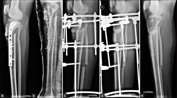
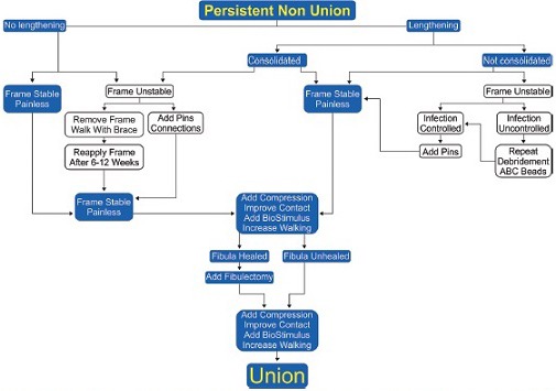
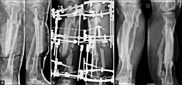
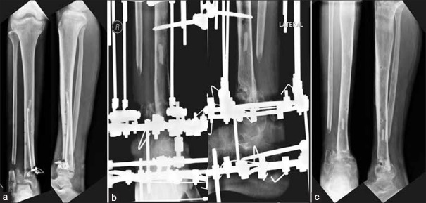
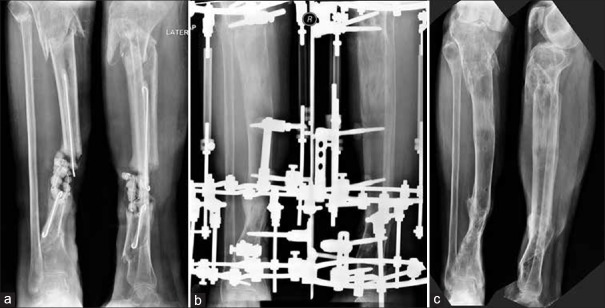
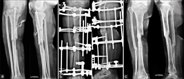
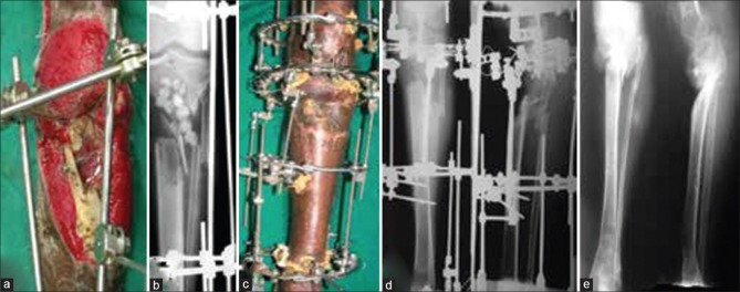
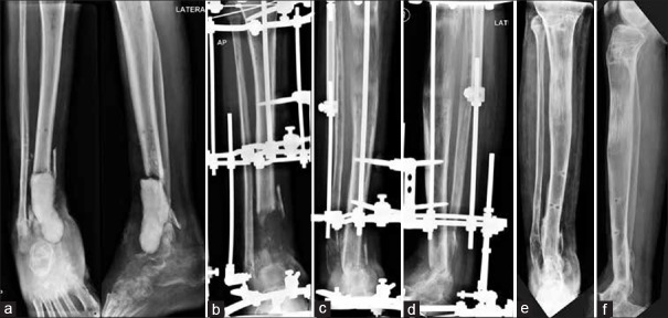
Infected nonunions of tibia pose many challenges to the treating surgeon and the patient. Challenges include recalcitrant infection, complex deformities, sclerotic bone ends, large bone gaps, shortening, and joint stiffness. They are easy to diagnose and difficult to treat. The ASAMI classification helps decide treatment. The nonunion severity score proposed by Calori measures many parameters to give a prognosis. The infection severity score uses simple clinical signs to grade severity of infection. This determines number of surgeries and allows choice of hardware, either external or internal for definitive treatment. Co-morbid factors such as smoking, diabetes, nonsteroidal anti-inflammatory drug use, and hypovitaminosis D influence the choice and duration of treatment. Thorough debridement is the mainstay of treatment. Removal of all necrotic bone and soft tissue is needed. Care is exercised in shaping bone ends. Internal fixation can help achieve union if infection was mild. Severe infections need external fixation use in a second stage. Compression at nonunion site achieves union. It can be combined with a corticotomy lengthening at a distant site for equalization. Soft tissue deficit has to be covered by flaps, either local or microvascular. Bone gaps are best filled with the reliable technique of bone transport. Regenerate bone may be formed proximally, distally, or at both sites. Acute compression can fill bone gaps and may need a fibular resection. Gradual reduction of bone gap happens with bone transport, without need for fibulectomy. When bone ends dock, union may be achieved by vertical or horizontal compression. Biological stimulus from iliac crest bone grafts, bone marrow aspirate injections, and platelet concentrates hasten union. Bone graft substitutes add volume to graft and help fill defects. Addition of rh-BMP-7 may help in healing albeit at a much higher cost. Regeneration may need stimulation and augmentation. Induced membrane technique is an alternative to bone transport to fill gaps. It needs large amounts of bone graft from iliac crest or femoral canal. This is an expensive method physiologically and economically. Infection can resorb the graft and cause failure of treatment. It can be done in select cases after thorough eradication of infection. Patience and perseverance are needed for successful resolution of infection and achieving union.
胫骨感染性骨不连给治疗医生和患者带来诸多挑战。挑战包括顽固性感染、复杂畸形、硬化骨端、大的骨缺损、短缩以及关节僵硬。它们易于诊断但难以治疗。ASAMI分类有助于确定治疗方案。Calori提出的骨不连严重程度评分可测量多个参数以给出预后情况。感染严重程度评分利用简单的临床体征对感染严重程度进行分级。这决定了手术次数,并允许选择用于确定性治疗的内固定或外固定器械。吸烟、糖尿病、使用非甾体抗炎药以及维生素D缺乏等合并因素会影响治疗的选择和持续时间。彻底清创是治疗的关键。需要清除所有坏死骨和软组织。修整骨端时需谨慎操作。如果感染较轻,内固定有助于实现骨愈合。严重感染则需在第二阶段使用外固定。骨不连部位的加压可实现骨愈合。可将其与远处的皮质切开延长术相结合以达到均衡。软组织缺损必须用局部或微血管皮瓣覆盖。骨缺损最好采用可靠的骨搬运技术填充。再生骨可在近端、远端或两端形成。急性加压可填充骨缺损,可能需要切除腓骨。骨搬运可逐渐缩小骨缺损,无需进行腓骨切除术。当骨端对接时,可通过垂直或水平加压实现骨愈合。来自髂嵴骨移植、骨髓抽吸注射和血小板浓缩物的生物刺激可加速骨愈合。骨移植替代物可增加移植骨的体积并有助于填充缺损。添加重组人骨形态发生蛋白-7可能有助于愈合,尽管成本要高得多。再生可能需要刺激和增强。诱导膜技术是替代骨搬运填充骨缺损的一种方法。它需要从髂嵴或股骨髓腔获取大量骨移植材料。这在生理和经济上都是一种昂贵的方法。感染可能会吸收移植骨并导致治疗失败。在彻底根除感染后,可在特定病例中采用该方法。成功解决感染并实现骨愈合需要耐心和毅力。