Düssmann Heiko, Perez-Alvarez Sergio, Anilkumar Ujval, Papkovsky Dmitri B, Prehn Jochen Hm
Department of Physiology and Medical Physics, Royal College of Surgeons in Ireland, 123 St. Stephen's Green, Dublin 2, Ireland.
Centre for Systems Medicine, Royal College of Surgeons in Ireland, 123 St. Stephen's Green, Dublin 2, Ireland.
Cell Death Dis. 2017 Jun 1;8(6):e2853. doi: 10.1038/cddis.2017.247.
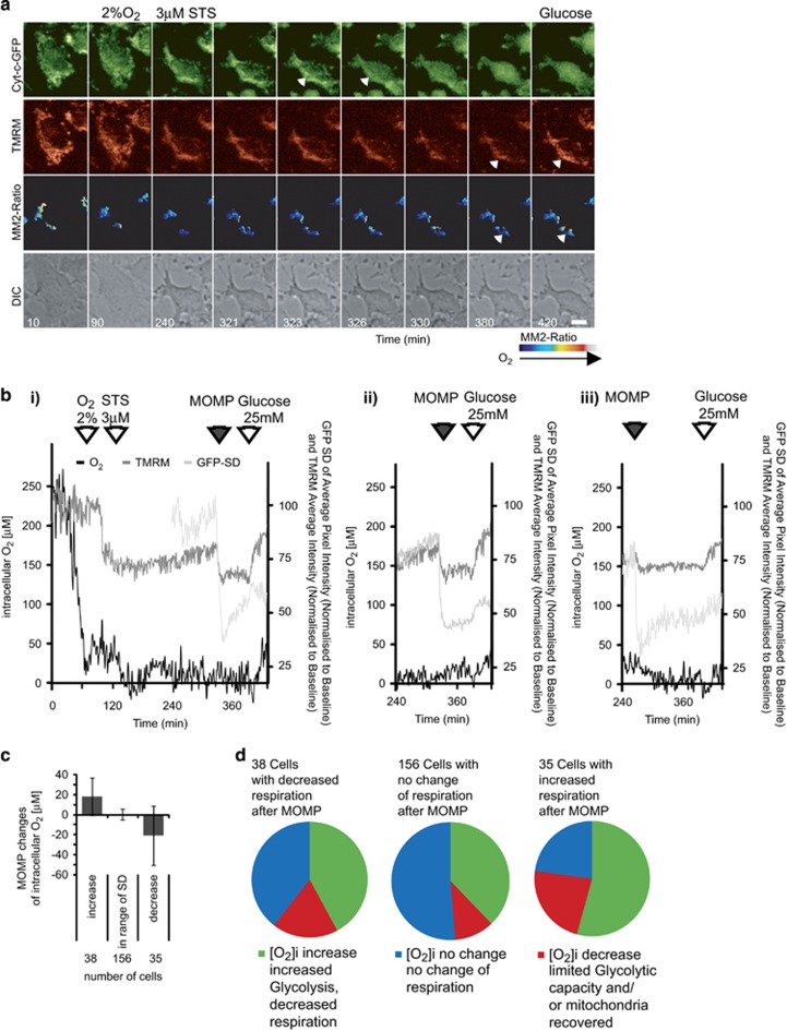
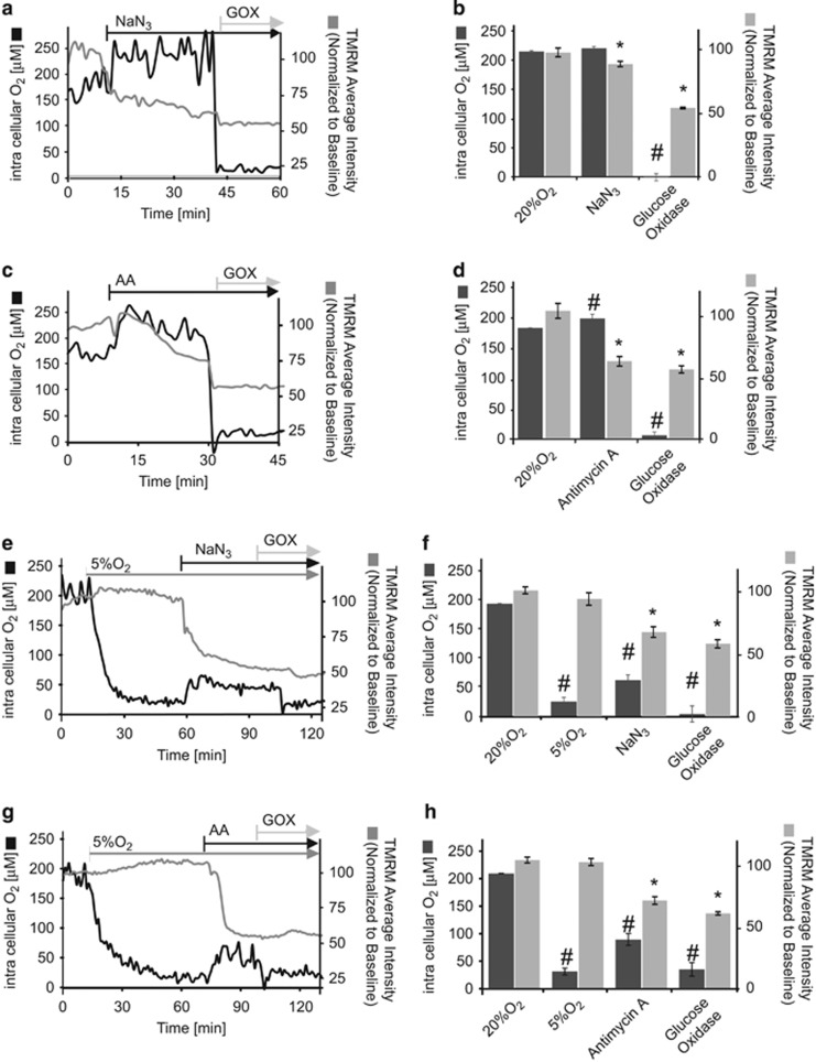
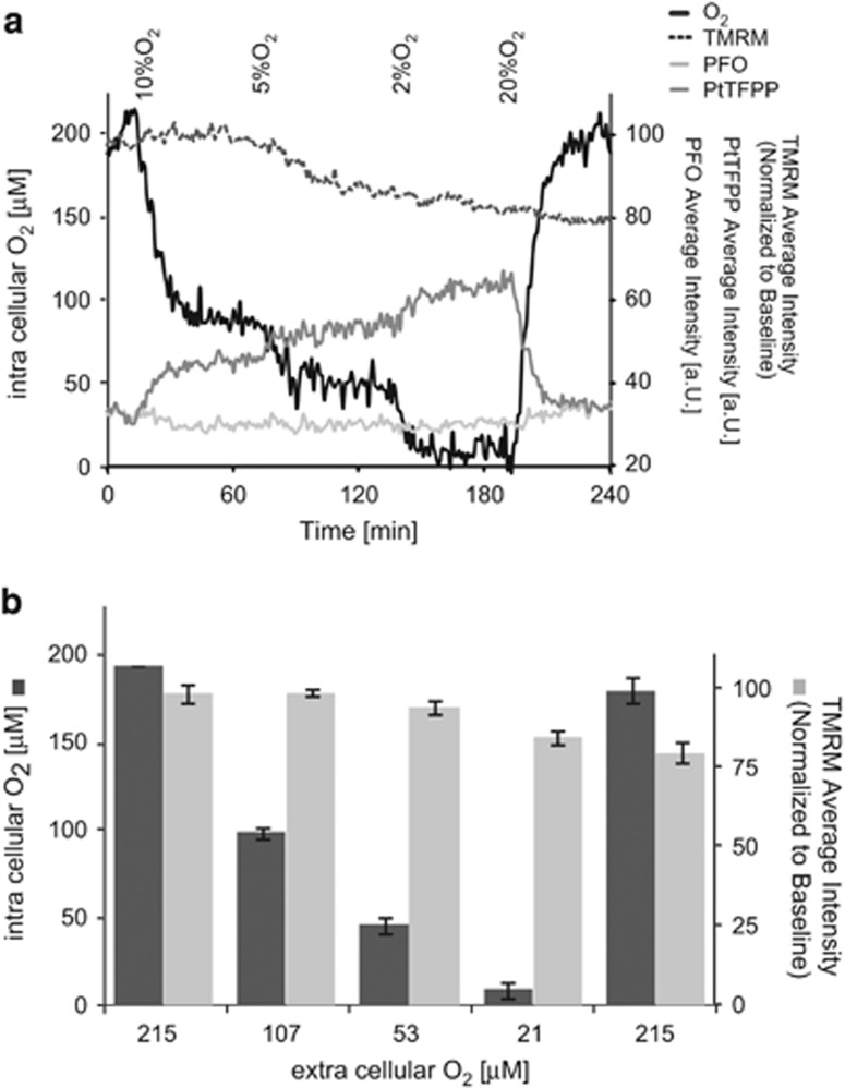
The detection of intracellular molecular oxygen (O) levels is important for understanding cell physiology, cell death, and drug effects, and has recently been improved with the development of oxygen-sensitive probes that are compatible with live cell time-lapse microscopy. We here provide a protocol for the use of the nanoparticle probe MitoImage-MM2 to monitor intracellular oxygen levels by confocal microscopy under baseline conditions, in response to mitochondrial toxins, and following mitochondrial cytochrome-c release. We demonstrate that the MitoImage-MM2 probe, which embeds Pt(II)-5,10,15,20-tetrakis-(2,3,4,5,6-pentafluorophenyl)-porphyrin as oxygen sensor and poly(9,9-dioctylfluorene) as an O-independent component, enables quantitative, ratiometric time-lapse imaging of intracellular O. Multiplexing with tetra-methyl-rhodamine-methyl ester in HeLa cervical cancer cells showed significant increases in intracellular O accompanied by strong mitochondrial depolarization when respiratory chain complexes III or IV were inhibited by Antimycin A or sodium azide, respectively, and when cells were maintained at 'physiological' tissue O levels (5% O). Multiplexing also allowed us to monitor intracellular O during the apoptotic signaling process of mitochondrial outer membrane permeabilization in HeLa expressing cytochrome-c-eGFP, and demonstrated that mitochondria post cytochrome-c release are able to retain their capacity to respire at physiological O despite a decrease in mitochondrial membrane potential.
细胞内分子氧(O)水平的检测对于理解细胞生理学、细胞死亡和药物作用至关重要,并且随着与活细胞延时显微镜兼容的氧敏感探针的发展,这一检测方法最近得到了改进。我们在此提供了一种使用纳米颗粒探针MitoImage-MM2的方案,用于在基线条件下、响应线粒体毒素以及线粒体细胞色素c释放后,通过共聚焦显微镜监测细胞内氧水平。我们证明,MitoImage-MM2探针嵌入了作为氧传感器的Pt(II)-5,10,15,20-四(2,3,4,5,6-五氟苯基)卟啉和作为与氧无关成分的聚(9,9-二辛基芴),能够对细胞内的O进行定量、比率延时成像。在HeLa宫颈癌细胞中与四甲基罗丹明甲酯进行多重检测显示,当呼吸链复合物III或IV分别被抗霉素A或叠氮化钠抑制时,以及当细胞维持在“生理”组织氧水平(5% O)时,细胞内O显著增加,同时伴有强烈的线粒体去极化。多重检测还使我们能够在表达细胞色素c-eGFP的HeLa细胞线粒体膜通透性改变的凋亡信号传导过程中监测细胞内O,并证明细胞色素c释放后的线粒体尽管线粒体膜电位降低,但仍能够在生理氧水平下保持呼吸能力。