Kurokawa Yoshika, Sone Hideko, Win-Shwe Tin-Tin, Zeng Yang, Kimura Hiroyuki, Koyama Yosuke, Yagi Yusuke, Matsui Yasuto, Yamazaki Masashi, Hirano Seishiro
Center for Health and Environmental Risk Research, National Institute for Environmental Studies, Tsukuba, Ibaraki.
Department of Analytical and Bioinorganic Chemistry, Kyoto Pharmaceutical University.
Int J Nanomedicine. 2017 May 24;12:3967-3975. doi: 10.2147/IJN.S125808. eCollection 2017.
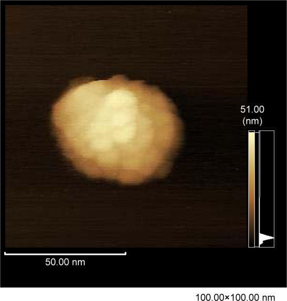
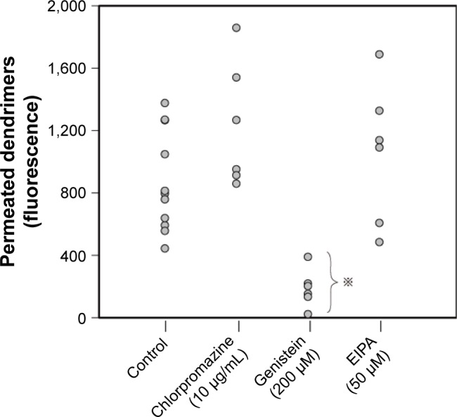
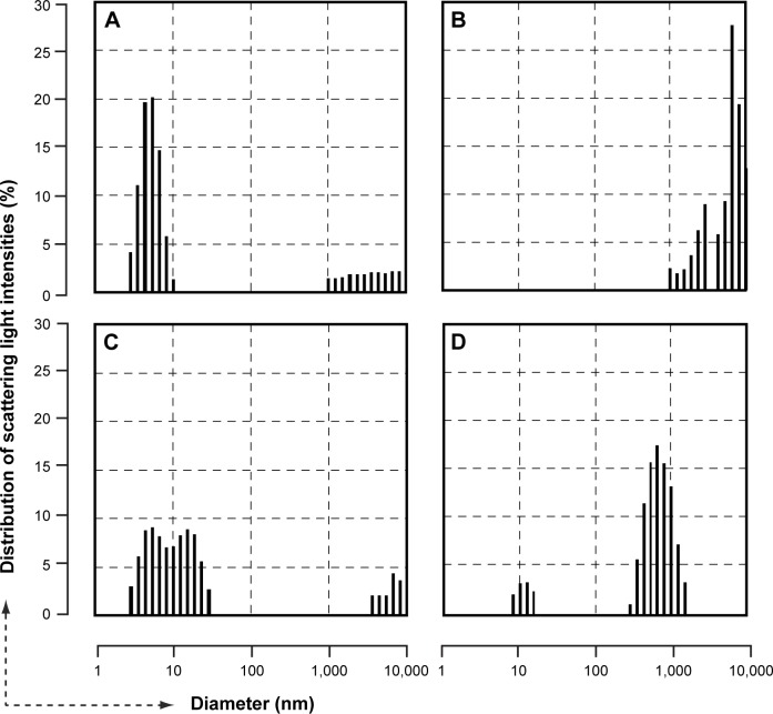
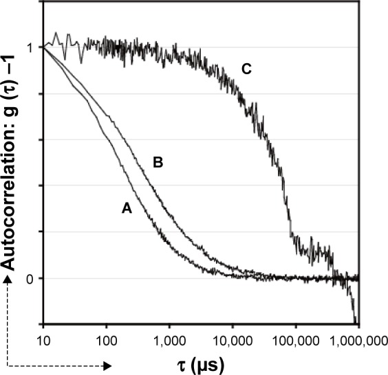
Dendrimers have been expected as excellent nanodevices for brain medication. An amine-terminated polyamidoamine dendrimer (PD), an unmodified plain type of PD, has the obvious disadvantage of cytotoxicity, but still serves as an attractive molecule because it easily adheres to the cell surface, facilitating easy cellular uptake. Single-photon emission computed tomographic imaging of a mouse following intravenous injection of a radiolabeled PD failed to reveal any signal in the intracranial region. Furthermore, examination of the permeability of PD particles across the blood-brain barrier (BBB) in vitro using a commercially available kit revealed poor permeability of the nanoparticles, which was suppressed by an inhibitor of caveolae-mediated endocytosis, but not by an inhibitor of macropinocytosis. Physicochemical analysis of the PD revealed that cationic PDs are likely to aggregate promptly upon mixing with body fluids and that this prompt aggregation is probably driven by non-Derjaguin-Landau- Verwey-Overbeek attractive forces originating from the surrounding divalent ions. Atomic force microscopy observation of a freshly cleaved mica plate soaked in dendrimer suspension (culture media) confirmed prompt aggregation. Our study revealed poor transfer of intravenously administered cationic PDs into the intracranial nervous tissue, and the results of our analysis suggested that this was largely attributable to the reduced BBB permeability arising from the propensity of the particles to promptly aggregate upon mixing with body fluids.
树枝状聚合物有望成为用于脑部治疗的出色纳米装置。胺基封端的聚酰胺胺树枝状聚合物(PD),一种未修饰的普通类型的PD,具有明显的细胞毒性缺点,但仍然是一种有吸引力的分子,因为它很容易附着在细胞表面,便于细胞摄取。对静脉注射放射性标记的PD后的小鼠进行单光子发射计算机断层扫描成像,未能在颅内区域发现任何信号。此外,使用市售试剂盒在体外检测PD颗粒穿过血脑屏障(BBB)的通透性,结果显示纳米颗粒的通透性较差,这种通透性受到小窝介导的内吞作用抑制剂的抑制,但不受巨胞饮作用抑制剂的抑制。对PD的物理化学分析表明,阳离子型PD与体液混合后可能会迅速聚集,这种迅速聚集可能是由周围二价离子产生的非德贾金-朗道-韦弗-奥弗贝克吸引力驱动的。原子力显微镜观察浸泡在树枝状聚合物悬浮液(培养基)中的新鲜劈开的云母片证实了迅速聚集。我们的研究表明静脉注射的阳离子型PD向颅内神经组织的转移较差,我们的分析结果表明,这在很大程度上归因于颗粒与体液混合后迅速聚集的倾向导致血脑屏障通透性降低。