Nikaki Alexandra, Angelidis George, Efthimiadou Roxani, Tsougos Ioannis, Valotassiou Varvara, Fountas Konstantinos, Prasopoulos Vasileios, Georgoulias Panagiotis
Department of Clinical Physiology, KHSHP, 20 Ahvenistontie Str., 13530, Hämeenlinna, Finland.
Department of Nuclear Medicine, University Hospital of Larissa, Mezourlo, 41110, Larissa, Greece.
Ann Nucl Med. 2017 Aug;31(7):495-505. doi: 10.1007/s12149-017-1183-2. Epub 2017 Jun 13.
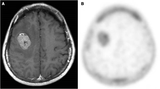
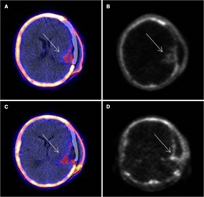
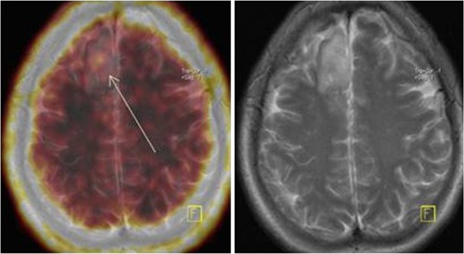
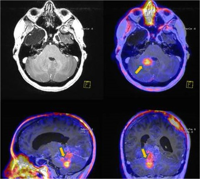
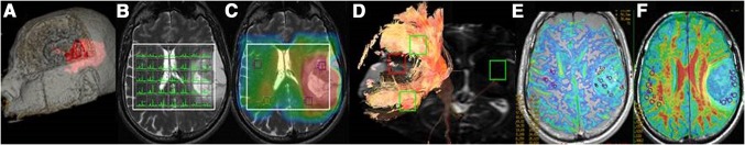
Brain neoplasms constitute a group of tumors with discrete differentiation grades, and therefore, course of disease and prognosis. Magnetic resonance imaging (MRI) remains the gold standard method for the investigation of central nervous system tumors. However, MRI suffers certain limitations, especially if radiation therapy or chemotherapy has been previously applied. On the other hand, given the development of newer radiopharmaceuticals, positron emission tomography (PET) aims to a better investigation of brain tumors, assisting in the clinical management of the patients. In the present review, the potential contribution of radiolabeled fluorothymidine (FLT) imaging for the evaluation of brain tumors will be discussed. In particular, we will present the role of FLT-PET imaging in the depiction of well and poorly differentiated lesions, the assessment of patient prognosis and treatment response, and the recognition of disease recurrence. Moreover, related semi-quantitative and kinetic parameters will be discussed.
脑肿瘤是一组具有不同分化程度的肿瘤,因此其病程和预后也各不相同。磁共振成像(MRI)仍然是中枢神经系统肿瘤检查的金标准方法。然而,MRI存在一定的局限性,尤其是在先前已进行过放射治疗或化疗的情况下。另一方面,鉴于新型放射性药物的发展,正电子发射断层扫描(PET)旨在更好地检查脑肿瘤,辅助患者的临床管理。在本综述中,将讨论放射性标记的氟代胸腺嘧啶核苷(FLT)成像在脑肿瘤评估中的潜在作用。特别是,我们将介绍FLT-PET成像在鉴别高分化和低分化病变、评估患者预后和治疗反应以及识别疾病复发方面的作用。此外,还将讨论相关的半定量和动力学参数。