• 文献检索
  • 文档翻译
  • 深度研究
  • 学术资讯
  • Suppr Zotero 插件Zotero 插件
  • 邀请有礼
  • 套餐&价格
  • 历史记录
应用&插件
Suppr Zotero 插件Zotero 插件浏览器插件Mac 客户端Windows 客户端微信小程序
定价
高级版会员购买积分包购买API积分包
服务
文献检索文档翻译深度研究API 文档MCP 服务
关于我们
关于 Suppr公司介绍联系我们用户协议隐私条款
关注我们

Suppr 超能文献

核心技术专利:CN118964589B侵权必究
粤ICP备2023148730 号-1Suppr @ 2026

文献检索

告别复杂PubMed语法,用中文像聊天一样搜索,搜遍4000万医学文献。AI智能推荐,让科研检索更轻松。

立即免费搜索

文件翻译

保留排版,准确专业,支持PDF/Word/PPT等文件格式,支持 12+语言互译。

免费翻译文档

深度研究

AI帮你快速写综述,25分钟生成高质量综述,智能提取关键信息,辅助科研写作。

立即免费体验

硒化镉/硫化锌量子点的细胞内运输在其后续毒性特征方面的作用。

The role of intracellular trafficking of CdSe/ZnS QDs on their consequent toxicity profile.

作者信息

Manshian Bella B, Martens Thomas F, Kantner Karsten, Braeckmans Kevin, De Smedt Stefaan C, Demeester Jo, Jenkins Gareth J S, Parak Wolfgang J, Pelaz Beatriz, Doak Shareen H, Himmelreich Uwe, Soenen Stefaan J

机构信息

Biomedical NMR Unit/MoSAIC, KU Leuven Campus Gasthuisberg, Herestraat 49, 3000, Louvain, Belgium.

Institute of Life Science, Swansea University Medical School, Singleton Park, Swansea, SA2 8PP, UK.

出版信息

J Nanobiotechnology. 2017 Jun 15;15(1):45. doi: 10.1186/s12951-017-0279-0.

DOI:10.1186/s12951-017-0279-0
PMID:28619032
原文链接:https://pmc.ncbi.nlm.nih.gov/articles/PMC5472855/
Abstract

BACKGROUND

Nanoparticle interactions with cellular membranes and the kinetics of their transport and localization are important determinants of their functionality and their biological consequences. Understanding these phenomena is fundamental for the translation of such NPs from in vitro to in vivo systems for bioimaging and medical applications. Two CdSe/ZnS quantum dots (QD) with differing surface functionality (NH or COOH moieties) were used here for investigating the intracellular uptake and transport kinetics of these QDs.

RESULTS

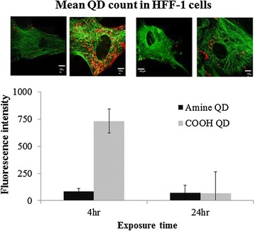
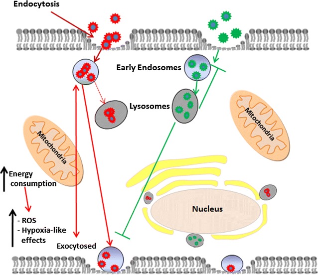
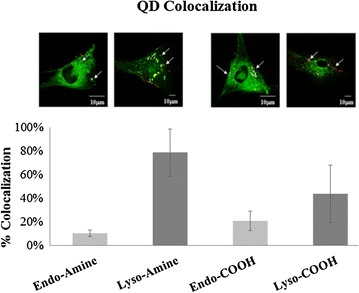
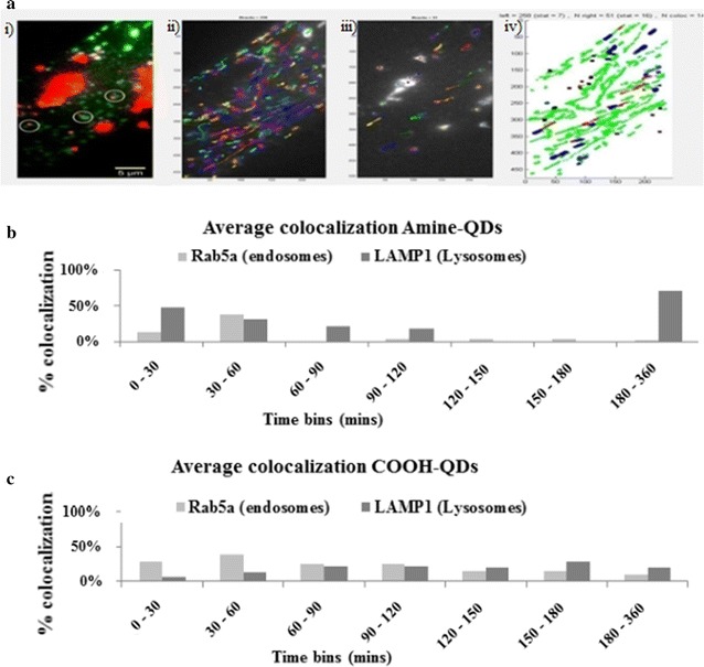
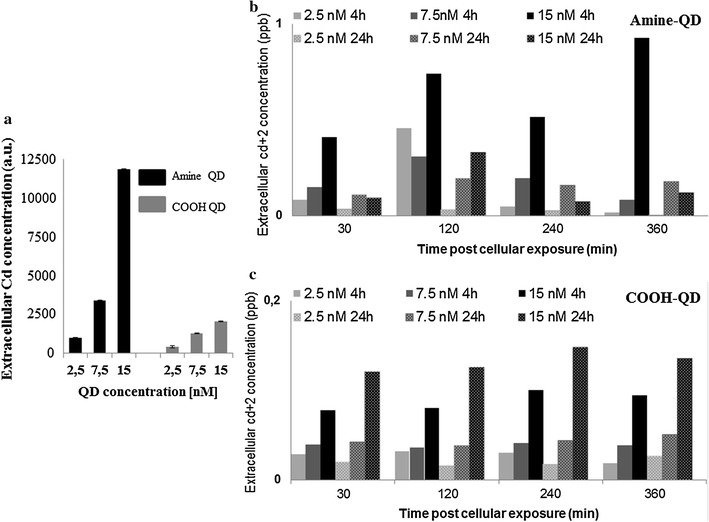
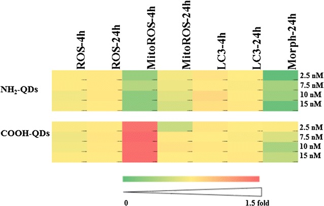
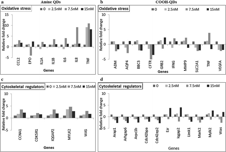
In water, the COOH- and NH-QDs were negatively and positively charged, respectively, while in serum-containing medium the NH-QDs were agglomerated, whereas the COOH-QDs remained dispersed. Though intracellular levels of NH- and COOH-QDs were very similar after 24 h exposure, COOH-QDs appeared to be continuously internalised and transported by endosomes and lysosomes, while NH-QDs mainly remained in the lysosomes. The results of (intra)cellular QD trafficking were correlated to their toxicity profiles investigating levels of reactive oxygen species (ROS), mitochondrial ROS, autophagy, changes to cellular morphology and alterations in genes involved in cellular stress, toxicity and cytoskeletal integrity. The continuous flux of COOH-QDs perhaps explains their higher toxicity compared to the NH-QDs, mainly resulting in mitochondrial ROS and cytoskeletal remodelling which are phenomena that occur early during cellular exposure.

CONCLUSIONS

Together, these data reveal that although cellular QD levels were similar after 24 h, differences in the nature and extent of their cellular trafficking resulted in differences in consequent gene alterations and toxicological effects.

摘要

背景

纳米颗粒与细胞膜的相互作用及其运输和定位动力学是其功能和生物学后果的重要决定因素。了解这些现象是将此类纳米颗粒从体外系统转化到体内系统用于生物成像和医学应用的基础。本文使用了两种具有不同表面功能(NH或COOH基团)的CdSe/ZnS量子点(QD)来研究这些量子点的细胞内摄取和运输动力学。

结果

在水中,COOH-QD和NH-QD分别带负电荷和正电荷,而在含血清的培养基中,NH-QD发生团聚,而COOH-QD仍保持分散状态。虽然暴露24小时后NH-QD和COOH-QD的细胞内水平非常相似,但COOH-QD似乎被内体和溶酶体持续内化和运输,而NH-QD主要留在溶酶体中。(细胞内)量子点运输的结果与其毒性特征相关,该毒性特征研究了活性氧(ROS)、线粒体ROS、自噬水平、细胞形态变化以及参与细胞应激、毒性和细胞骨架完整性的基因改变。COOH-QD的持续通量可能解释了其与NH-QD相比更高的毒性,主要导致线粒体ROS和细胞骨架重塑,这些是细胞暴露早期发生的现象。

结论

总之,这些数据表明,尽管24小时后细胞内量子点水平相似,但它们在细胞运输的性质和程度上的差异导致了随后基因改变和毒理学效应的差异。

https://cdn.ncbi.nlm.nih.gov/pmc/blobs/7990/5472855/d671ffaba6a5/12951_2017_279_Fig7_HTML.jpg
https://cdn.ncbi.nlm.nih.gov/pmc/blobs/7990/5472855/75ef1a40e79a/12951_2017_279_Fig1_HTML.jpg
https://cdn.ncbi.nlm.nih.gov/pmc/blobs/7990/5472855/6b4dd237d7be/12951_2017_279_Fig2_HTML.jpg
https://cdn.ncbi.nlm.nih.gov/pmc/blobs/7990/5472855/37d5a6c13f38/12951_2017_279_Fig3_HTML.jpg
https://cdn.ncbi.nlm.nih.gov/pmc/blobs/7990/5472855/50fd16bb5c94/12951_2017_279_Fig4_HTML.jpg
https://cdn.ncbi.nlm.nih.gov/pmc/blobs/7990/5472855/a87a4aa04191/12951_2017_279_Fig5_HTML.jpg
https://cdn.ncbi.nlm.nih.gov/pmc/blobs/7990/5472855/bddbe9f55e91/12951_2017_279_Fig6_HTML.jpg
https://cdn.ncbi.nlm.nih.gov/pmc/blobs/7990/5472855/d671ffaba6a5/12951_2017_279_Fig7_HTML.jpg

相似文献

1
The role of intracellular trafficking of CdSe/ZnS QDs on their consequent toxicity profile.硒化镉/硫化锌量子点的细胞内运输在其后续毒性特征方面的作用。
J Nanobiotechnology. 2017 Jun 15;15(1):45. doi: 10.1186/s12951-017-0279-0.
2
The cytotoxicities in prokaryote and eukaryote varied for CdSe and CdSe/ZnS quantum dots and differed from cadmium ions.CdSe 和 CdSe/ZnS 量子点对原核生物和真核生物的细胞毒性与镉离子不同。
Ecotoxicol Environ Saf. 2019 Oct 15;181:336-344. doi: 10.1016/j.ecoenv.2019.06.027. Epub 2019 Jun 13.
3
Metallomics Study of CdSe/ZnS Quantum Dots in HepG2 Cells.CdSe/ZnS 量子点在 HepG2 细胞中的金属组学研究。
ACS Nano. 2015 Oct 27;9(10):10324-34. doi: 10.1021/acsnano.5b04365. Epub 2015 Sep 30.
4
Transcriptome Profile Alteration with Cadmium Selenide/Zinc Sulfide Quantum Dots in .硒化镉/硫化锌量子点对 . 的转录组谱改变
Biomolecules. 2019 Oct 25;9(11):653. doi: 10.3390/biom9110653.
5
Cellular uptake, elimination and toxicity of CdSe/ZnS quantum dots in HepG2 cells.CdSe/ZnS 量子点在 HepG2 细胞中的细胞摄取、消除和毒性。
Biomaterials. 2013 Dec;34(37):9545-58. doi: 10.1016/j.biomaterials.2013.08.038. Epub 2013 Sep 5.
6
Enantioselective cellular uptake of chiral semiconductor nanocrystals.手性半导体纳米晶体的对映选择性细胞摄取。
Nanotechnology. 2016 Feb 19;27(7):075102. doi: 10.1088/0957-4484/27/7/075102. Epub 2016 Jan 19.
7
The interactions between CdSe quantum dots and yeast Saccharomyces cerevisiae: adhesion of quantum dots to the cell surface and the protection effect of ZnS shell.硒化镉量子点与酿酒酵母的相互作用:量子点与细胞表面的黏附及硫化锌壳的保护作用。
Chemosphere. 2014 Oct;112:92-9. doi: 10.1016/j.chemosphere.2014.03.071. Epub 2014 Apr 21.
8
Evaluation of quantum dot cytotoxicity: interpretation of nanoparticle concentrations versus intracellular nanoparticle numbers.量子点细胞毒性评估:纳米颗粒浓度与细胞内纳米颗粒数量的解读
Nanotoxicology. 2016 Nov;10(9):1318-28. doi: 10.1080/17435390.2016.1210691. Epub 2016 Jul 28.
9
Toxicological effects of CdSe/ZnS quantum dots on marine planktonic organisms.CdSe/ZnS量子点对海洋浮游生物的毒理学效应
Ecotoxicol Environ Saf. 2016 Jan;123:26-31. doi: 10.1016/j.ecoenv.2015.09.020. Epub 2015 Sep 26.
10
CdSe/ZnS quantum dots exhibited nephrotoxicity through mediating oxidative damage and inflammatory response.硒化镉/硫化锌量子点通过介导氧化损伤和炎症反应表现出肾毒性。
Aging (Albany NY). 2020 Nov 16;13(8):12194-12206. doi: 10.18632/aging.103774.

引用本文的文献

1
Surface Charge Affects the Intracellular Fate and Clearance Dynamics of CdSe/ZnS Quantum Dots in Macrophages.表面电荷影响巨噬细胞中CdSe/ZnS量子点的细胞内命运和清除动力学。
Nanomaterials (Basel). 2025 Aug 3;15(15):1189. doi: 10.3390/nano15151189.
2
Applicability of Quantum Dots in Breast Cancer Diagnostic and Therapeutic Modalities-A State-of-the-Art Review.量子点在乳腺癌诊断与治疗方式中的适用性——最新综述
Nanomaterials (Basel). 2024 Aug 31;14(17):1424. doi: 10.3390/nano14171424.
3
A Comparison of Common Quantum Dot Alternatives to Cadmium-Based Quantum Dots on the Basis of Liver Cytotoxicity.

本文引用的文献

1
Probing the Cytotoxicity Of Semiconductor Quantum Dots.探究半导体量子点的细胞毒性
Nano Lett. 2004 Jan 1;4(1):11-18. doi: 10.1021/nl0347334. Epub 2003 Dec 10.
2
Optimizing conditions for labeling of mesenchymal stromal cells (MSCs) with gold nanoparticles: a prerequisite for in vivo tracking of MSCs.优化用金纳米颗粒标记间充质基质细胞(MSCs)的条件:MSCs体内追踪的先决条件。
J Nanobiotechnology. 2017 Mar 29;15(1):24. doi: 10.1186/s12951-017-0258-5.
3
Dissecting common and divergent molecular pathways elicited by CdSe/ZnS quantum dots in freshwater and marine sentinel invertebrates.
基于肝细胞毒性对镉基量子点常见替代物的比较
Nanomaterials (Basel). 2024 Jun 25;14(13):1086. doi: 10.3390/nano14131086.
4
Cytotoxicity of Quantum Dots in Receptor-Mediated Endocytic and Pinocytic Pathways in Yeast.量子点在酵母中受体介导的内吞和胞饮途径中的细胞毒性。
Int J Mol Sci. 2024 Apr 26;25(9):4714. doi: 10.3390/ijms25094714.
5
A Review of in vivo Toxicity of Quantum Dots in Animal Models.量子点在动物模型体内毒性的综述。
Int J Nanomedicine. 2023 Dec 29;18:8143-8168. doi: 10.2147/IJN.S434842. eCollection 2023.
6
Recent Breakthroughs in Using Quantum Dots for Cancer Imaging and Drug Delivery Purposes.量子点在癌症成像与药物递送方面的近期突破
Nanomaterials (Basel). 2023 Sep 15;13(18):2566. doi: 10.3390/nano13182566.
7
Current Advances in the Biomedical Applications of Quantum Dots: Promises and Challenges.当前量子点在生物医学应用中的进展:前景与挑战。
Int J Mol Sci. 2023 Aug 11;24(16):12682. doi: 10.3390/ijms241612682.
8
Apoptotic pathway protein expression variance in metal oxide and quantum dot treated HeLa cells.金属氧化物和量子点处理的HeLa细胞中凋亡途径蛋白表达的差异
MicroPubl Biol. 2023 Jul 18;2023. doi: 10.17912/micropub.biology.000801. eCollection 2023.
9
Exocytosis of Nanoparticles: A Comprehensive Review.纳米颗粒的胞吐作用:综述
Nanomaterials (Basel). 2023 Jul 30;13(15):2215. doi: 10.3390/nano13152215.
10
Current applications of nanomaterials in urinary system tumors.纳米材料在泌尿系统肿瘤中的当前应用。
Front Bioeng Biotechnol. 2023 Feb 20;11:1111977. doi: 10.3389/fbioe.2023.1111977. eCollection 2023.
剖析CdSe/ZnS量子点在淡水和海洋哨兵无脊椎动物中引发的共同和不同分子途径。
Nanotoxicology. 2017 Mar;11(2):289-303. doi: 10.1080/17435390.2017.1295111.
4
Basic Physicochemical Properties of Polyethylene Glycol Coated Gold Nanoparticles that Determine Their Interaction with Cells.聚乙二醇包覆金纳米粒子的基本物理化学性质决定了其与细胞的相互作用。
Angew Chem Int Ed Engl. 2016 Apr 25;55(18):5483-7. doi: 10.1002/anie.201511733. Epub 2016 Mar 29.
5
Efficient Subcellular Targeting to the Cell Nucleus of Quantum Dots Densely Decorated with a Nuclear Localization Sequence Peptide.高效亚细胞靶向至密集修饰有核定位序列肽的量子点的细胞核。
ACS Appl Mater Interfaces. 2016 Jan 27;8(3):2001-9. doi: 10.1021/acsami.5b10295. Epub 2016 Jan 14.
6
Charge and agglomeration dependent in vitro uptake and cytotoxicity of zinc oxide nanoparticles.氧化锌纳米颗粒的电荷和团聚对其体外摄取及细胞毒性的影响
J Inorg Biochem. 2015 Dec;153:334-338. doi: 10.1016/j.jinorgbio.2015.08.029. Epub 2015 Sep 3.
7
High-Content Imaging and Gene Expression Approaches To Unravel the Effect of Surface Functionality on Cellular Interactions of Silver Nanoparticles.高通量成像和基因表达方法揭示表面功能对银纳米粒子与细胞相互作用的影响。
ACS Nano. 2015 Oct 27;9(10):10431-44. doi: 10.1021/acsnano.5b04661. Epub 2015 Sep 3.
8
Where Are We Heading in Nanotechnology Environmental Health and Safety and Materials Characterization?纳米技术的环境健康与安全以及材料表征将何去何从?
ACS Nano. 2015 Jun 23;9(6):5627-30. doi: 10.1021/acsnano.5b03496.
9
(Intra)cellular stability of inorganic nanoparticles: effects on cytotoxicity, particle functionality, and biomedical applications.无机纳米颗粒的(细胞)内稳定性:对细胞毒性、颗粒功能及生物医学应用的影响
Chem Rev. 2015 Mar 11;115(5):2109-35. doi: 10.1021/cr400714j. Epub 2015 Feb 2.
10
Understanding nanoparticle cellular entry: A physicochemical perspective.理解纳米颗粒的细胞内进入:物理化学视角。
Adv Colloid Interface Sci. 2015 Apr;218:48-68. doi: 10.1016/j.cis.2015.01.007. Epub 2015 Feb 12.