Febbraro Fabia, Svenningsen Katrine, Tran Thao Phuong, Wiborg Ove
Danish Research Institute of Translational Neuroscience (DANDRITE) Aarhus University, Aarhus C, Denmark.
Focused Research Unit for Molecular Diagnostic and Clinical Research IRS-Center Sonderjylland, Laboratory Center, Hospital of Southern Jutland, Åbenrå, Denmark.
PLoS One. 2017 Jun 16;12(6):e0179434. doi: 10.1371/journal.pone.0179434. eCollection 2017.
Stress and stressful life events have repeatedly been shown as causally related to depression. The Chronic Mild Stress rat model is a valid model of stress-induced depression. Like humans, rats display great heterogeneity in their response to stress and adversity. Hence some individuals are stress-sensitive and prone to develop depression-like behaviour in response to modest stressors, while others are stress-resilient and remain essentially symptom free.
Compared to the large body of research, which describes stress-induced maladaptive neurobiological changes, relatively little attention has been devoted to understand resiliency to stress. The aim of the present study was to identify changes in neuronal activity, associated with stress-resilient and stress-susceptible chronic mild stress endophenotypes, by examining c-Fos expression in 13 different brain areas. Changes in c-Fos expression have been reported as associated to stressful conditions.
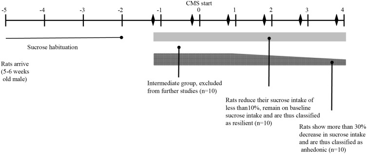
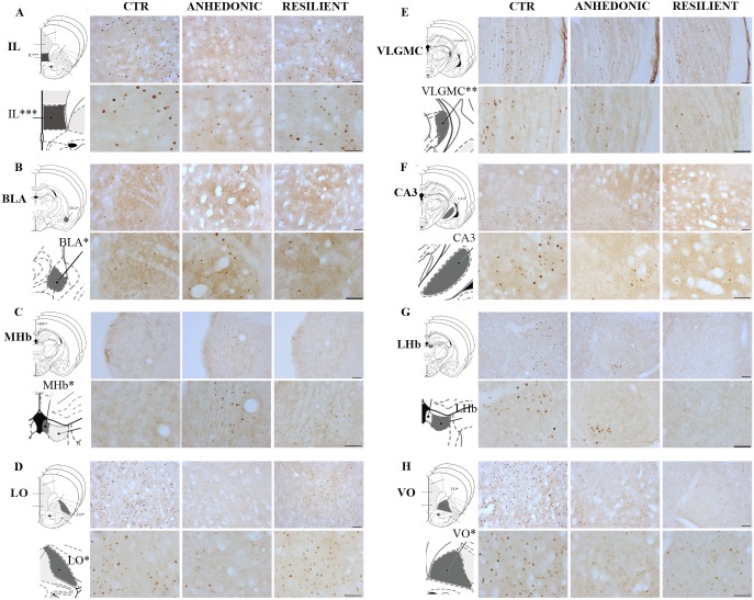
Stress-induced modulation of neuronal activation patterns in response to the chronic mild stress paradigm was mapped using the immediate early gene expression c-Fos as a marker. Quantification of the c-Fos-like immunoreactivity responses was done by semi-automated profile counting procedures and design-based stereology.
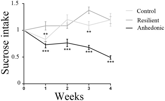
Exposure to chronic mild stress significantly altered c-Fos expression in a total of 6 out of 13 investigated areas. Chronic mild stress was found to suppress the c-Fos response within the magnocellular ventral lateral geniculate nucleus of both stress subgroups. In the the lateral and ventral orbital cortices of stress-resilient rats, the c-Fos like immunoreactivity response was also repressed by stress exposure. On the contrary the c-Fos response within the amygdala, medial habenula, and infralimbic cortex was increased selectively for the stress-susceptible rats.
The study was initiated to characterize neuronal substrates associated with stress-coping mechanisms. Six areas, all of which represents limbic structures, were found to be sensitive to stress exposure. The effects within these areas associate to the hedonic status of the rats. Hence, these areas might be associated to stress-coping mechanisms underlying the chronic mild stress induced segregation into stress-susceptible and stress-resilient endophenotypes.
压力和应激性生活事件一再被证明与抑郁症存在因果关系。慢性轻度应激大鼠模型是应激诱导抑郁症的有效模型。与人类一样,大鼠在对应激和逆境的反应中表现出很大的异质性。因此,一些个体对应激敏感,在面对适度应激源时容易出现类似抑郁的行为,而另一些个体则具有应激恢复力,基本无症状。
与大量描述应激诱导的适应不良神经生物学变化的研究相比,相对较少关注对应激恢复力的理解。本研究的目的是通过检查13个不同脑区的c-Fos表达,确定与应激恢复力和应激易感性慢性轻度应激内表型相关的神经元活动变化。据报道,c-Fos表达的变化与应激状态有关。
使用即刻早期基因表达c-Fos作为标记,绘制应激诱导的神经元激活模式对慢性轻度应激范式的调节图。通过半自动轮廓计数程序和基于设计的体视学对c-Fos样免疫反应进行定量。
暴露于慢性轻度应激后,13个研究区域中有6个区域的c-Fos表达发生了显著变化。发现慢性轻度应激会抑制两个应激亚组的大细胞腹侧外侧膝状核内的c-Fos反应。在应激恢复力强的大鼠的外侧和腹侧眶皮质中,应激暴露也会抑制c-Fos样免疫反应。相反,杏仁核、内侧缰核和边缘下皮质内的c-Fos反应在应激易感性大鼠中选择性增加。
本研究旨在表征与应激应对机制相关的神经元底物。发现有六个区域,均代表边缘结构,对应激暴露敏感。这些区域内的效应与大鼠的享乐状态相关。因此,这些区域可能与慢性轻度应激诱导分为应激易感性和应激恢复力内表型的应激应对机制有关。