Ma Jie, Li Zhi-Yuan, Liang Xiao-Peng, Guo Cai-Xia, Lu Pei-Pei, Ma Li-Hong
Department of Traditional Chinese Medicine, State Key Laboratory of Cardiovascular Disease, Fuwai Hospital, National Center for Cardiovascular Diseases, Chinese Academy of Medical Sciences and Peking Union Medical College, Beijing, China.
J Geriatr Cardiol. 2017 May;14(5):301-307. doi: 10.11909/j.issn.1671-5411.2017.05.005.
Recent clinical and experimental studies have confirmed the effects of Xinfuli Granule (XG), a compound Chinese medicine in the prevention and treatment of heart failure (HF). This study aimed to investigate the effects and the mechanisms of XG on ventricular reconstruction in rats with acute myocardial infarction (AMI).
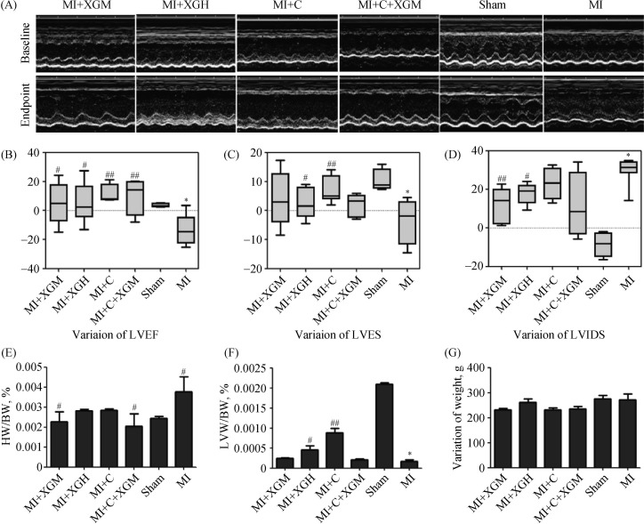
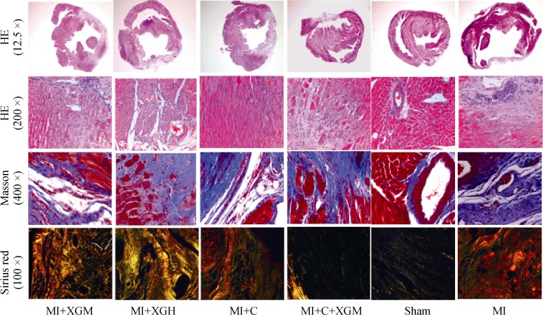
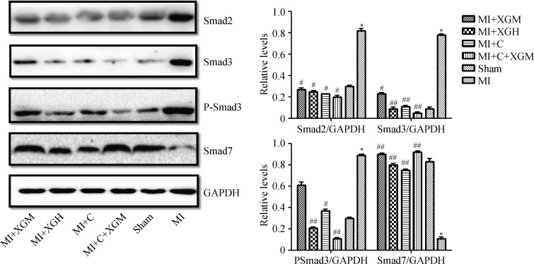
Sprague-Dawley rats were subjected to left anterior descending branch ligation. The rats that survived 24 h were randomly assigned to five groups: medium-dose of XG group (MI+XGM), high-dose of XG group (MI+XGH), carvedilol group (MI+C), medium-dose of XG + carvedilol group (MI+C+XGM). Fourteen rats underwent identical surgical procedures without artery ligation, serving as sham controls. At 28 days, left ventricular weight to body weight (LVW/BW) and heart weight to body weight (HW/BW) were calculated; left ventricular ejection fraction (LVEF), left ventricular shortening fraction (LVFS), left ventricular internal diameter at systole (LVIDS) were measured by ultrasound; HE staining, Masson staining, and Sirius red staining were used to assess the myocardial pathological and physiological changes as well as myocardial fibrosis area and non-infarct zone I/III collagen ratio. Expression of Smad3 were detected and analyzed by Western blot, immunohistochemistry and immunofluorescence. P-Smad3, Smad2 and Smad7 in the TGF-β/Smads signaling pathway were also analyzed by Western blot.
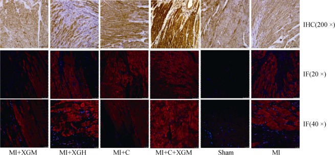
The LVIDS ( < 0.01), HW/BW ( < 0.05), type I/III collagen ratio ( < 0.01) and myocardial collagen ( < 0.01) decreased significantly while the LVW/BW, LVFS ( < 0.05) increased significantly in MI+XGM group as compared with those in other groups. The expression of key signal molecules of the TGF-β/Smads signaling pathway, including Smad3, P-Smad3 and Smad2 protein were decreased, while the expression of Smad7 increased in both XG and carvedilol treatment groups as compared to those of the MI group (all < 0.01). Immunohistochemistry and immunofluorescence further confirmed the down-regulated Smad3 expression.
XG can improve ventricular reconstruction and inhibit myocardial fibrosis in rats with AMI by regulating TGF-β/Smads signaling pathway.
近期的临床和实验研究证实了复方中药心复力颗粒(XG)在预防和治疗心力衰竭(HF)方面的作用。本研究旨在探讨XG对急性心肌梗死(AMI)大鼠心室重构的影响及其机制。
将Sprague-Dawley大鼠进行左前降支结扎。存活24小时的大鼠随机分为五组:XG中剂量组(MI+XGM)、XG高剂量组(MI+XGH)、卡维地洛组(MI+C)、XG中剂量+卡维地洛组(MI+C+XGM)。14只大鼠接受相同的手术操作但不结扎动脉,作为假手术对照组。在第28天,计算左心室重量与体重之比(LVW/BW)和心脏重量与体重之比(HW/BW);通过超声测量左心室射血分数(LVEF)、左心室短轴缩短率(LVFS)、收缩末期左心室内径(LVIDS);采用苏木精-伊红(HE)染色、Masson染色和天狼星红染色评估心肌病理和生理变化以及心肌纤维化面积和非梗死区I/III型胶原比例。通过蛋白质免疫印迹法、免疫组织化学和免疫荧光检测并分析Smad3的表达。还通过蛋白质免疫印迹法分析TGF-β/Smads信号通路中的P-Smad3、Smad2和Smad7。
与其他组相比,MI+XGM组的LVIDS(<0.01)、HW/BW(<0.05)、I/III型胶原比例(<0.01)和心肌胶原(<0.01)显著降低,而LVW/BW、LVFS(<0.05)显著增加。与MI组相比,XG和卡维地洛治疗组中TGF-β/Smads信号通路关键信号分子Smad3、P-Smad3和Smad2蛋白的表达降低,而Smad7的表达增加(均<0.01)。免疫组织化学和免疫荧光进一步证实了Smad3表达下调。
XG可通过调节TGF-β/Smads信号通路改善AMI大鼠的心室重构并抑制心肌纤维化。