The Key Laboratory of Cardiovascular Remodeling and Function Research, Chinese Ministry of Education and Chinese Ministry of Health, The State and Shandong Province Joint Key Laboratory of Translational Cardiovascular Medicine, Department of Cardiology, Qilu Hospital of Shandong University, Jinan, Shandong, China.
Department of General Practice, Qilu Hospital of Shandong University, Jinan, Shandong, China.
J Cell Mol Med. 2017 Dec;21(12):3298-3308. doi: 10.1111/jcmm.13233. Epub 2017 Jun 19.
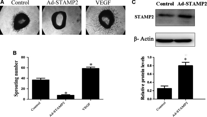
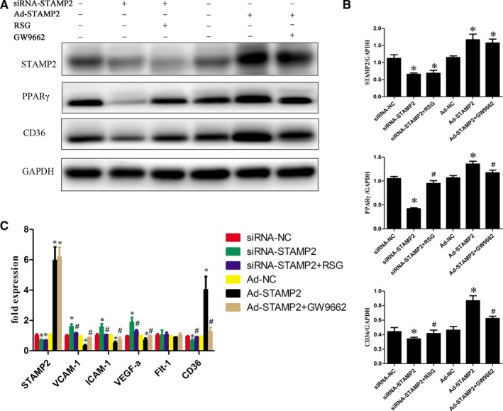
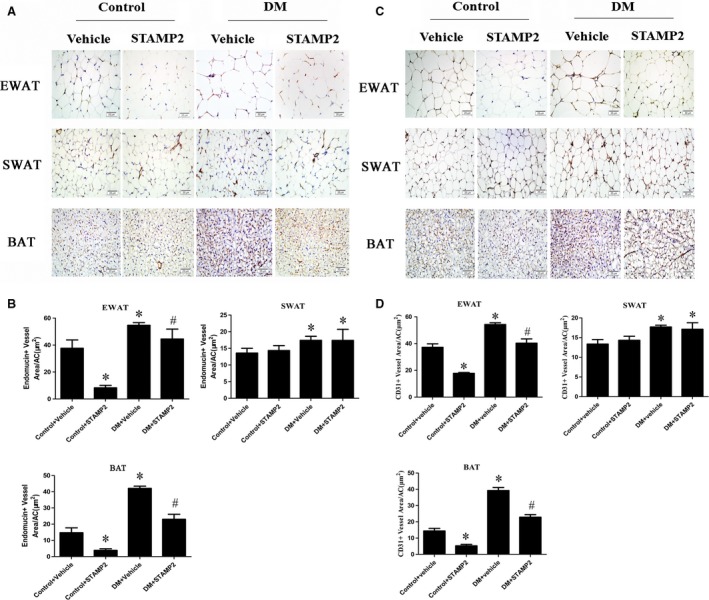
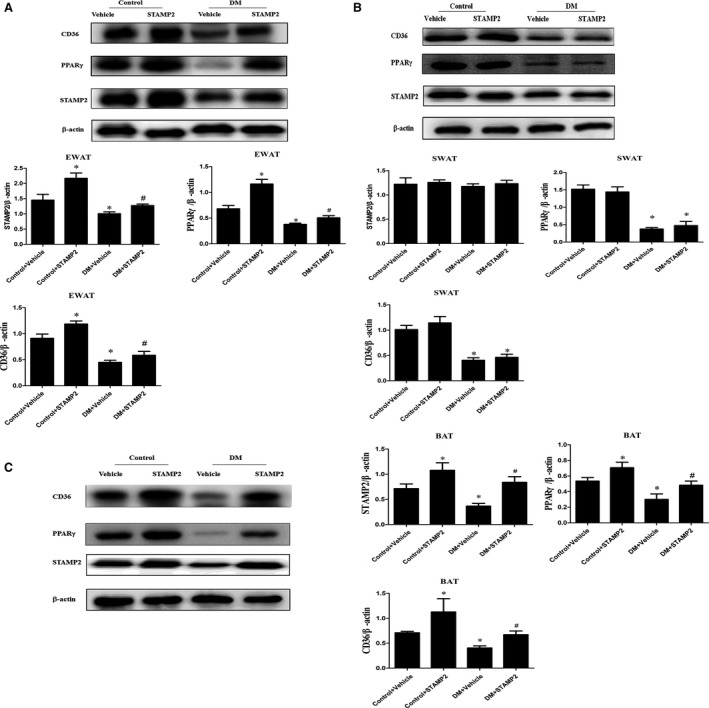
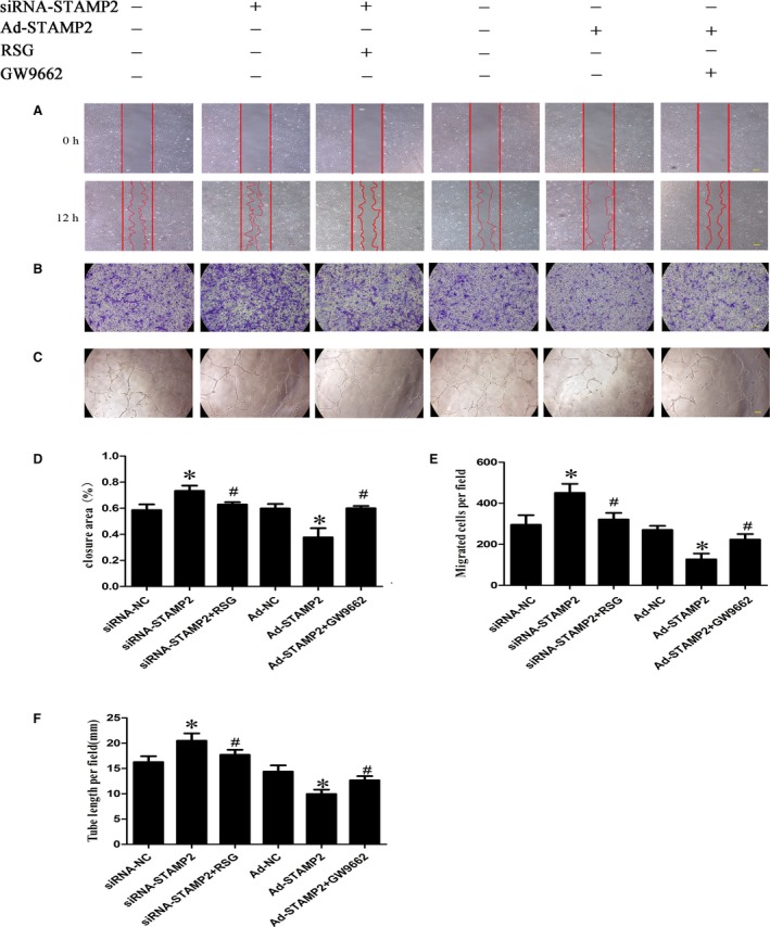
The aim of this study was to investigate whether overexpression of STAMP2 improves insulin resistance by regulating angiogenesis in adipose tissues. The characteristics of diabetic mice were measured by serial metabolite and pathology tests. Samples were obtained from epididymal, subcutaneous and brown adipose tissues. Histological and morphological analysis demonstrated that STAMP2 gene overexpression reduced adipocyte size, angiogenesis in epididymal and brown adipose tissues. On aortic ring assay, microvessels sprouting from aortas were significantly inhibited after STAMP2 gene overexpression. The cellular effect of STAMP2 on angiogenesis was explored in human umbilical vein endothelial cells (HUVECs) model. Correlation of STAMP2 and angiogenesis was validated by Ad-STAMP2 transfection and STAMP2 siRNA inhibition. In vitro, overexpression of STAMP2 significantly inhibited endothelial cell migration, tube formation. The effects of Ad-STAMP2 transfection on HUVECs were abolished by treatment with PPARγ antagonist GW9662 (2.5 μM), and the roles of STAMP2 siRNA on HUVECs were also reversed by treatment with PPARγ agonist rosiglitazone (RSG) (0.1 mM). RT-PCR indicated that STAMP2 could regulate levels of adhesion molecules, vascular endothelial growth factor A and CD36. The expression of PPARγ and CD36 was decreased when STAMP2 was inhibited by siRNA, while PPARγ and CD36 were highly expressed after overexpression of STAMP2. Our results suggested that STAMP2 gene overexpression may improve insulin resistance via attenuating angiogenesis in epididymal and brown adipose tissues through the PPARγ/CD36 signalling pathway.
本研究旨在探讨 STAMP2 的过表达是否通过调节脂肪组织中的血管生成来改善胰岛素抵抗。通过连续的代谢物和病理学测试来测量糖尿病小鼠的特征。从附睾、皮下和棕色脂肪组织中获取样本。组织学和形态学分析表明,STAMP2 基因过表达可减小脂肪细胞大小、附睾和棕色脂肪组织中的血管生成。在主动脉环测定中,STAMP2 基因过表达后,从主动脉中长出的微血管明显受到抑制。在人脐静脉内皮细胞 (HUVEC) 模型中探讨了 STAMP2 对血管生成的细胞作用。通过 Ad-STAMP2 转染和 STAMP2 siRNA 抑制验证了 STAMP2 与血管生成的相关性。在体外,STAMP2 的过表达显著抑制内皮细胞迁移、管形成。用 PPARγ 拮抗剂 GW9662(2.5 μM)处理可消除 Ad-STAMP2 转染对 HUVECs 的影响,用 PPARγ 激动剂罗格列酮(RSG)(0.1 mM)处理也可逆转 STAMP2 siRNA 对 HUVECs 的作用。RT-PCR 表明,STAMP2 可调节黏附分子、血管内皮生长因子 A 和 CD36 的水平。用 siRNA 抑制 STAMP2 时,PPARγ 和 CD36 的表达减少,而过表达 STAMP2 后,PPARγ 和 CD36 的表达增加。我们的结果表明,STAMP2 基因过表达可能通过 PPARγ/CD36 信号通路减弱附睾和棕色脂肪组织中的血管生成来改善胰岛素抵抗。