Molecular Science and Biomedicine Laboratory, State Key Laboratory of Chemo/Bio-Sensing and Chemometrics, College of Chemistry and Chemical Engineering, College of Biology, Hunan University, Changsha 410082, China.
Faculty of Science and Technology, University of Macau, Av. da Universidade, Taipa 999078, Macau.
Nat Commun. 2017 Jun 15;8:15653. doi: 10.1038/ncomms15653.
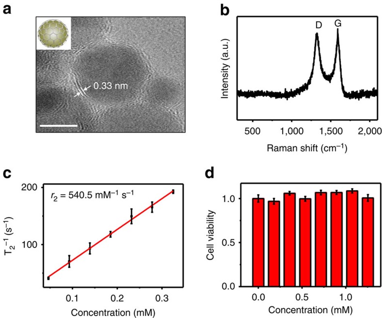
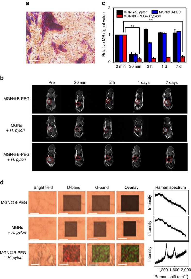
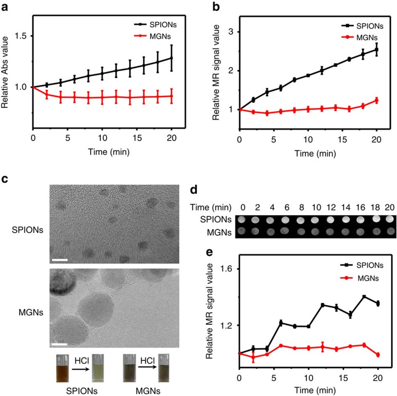
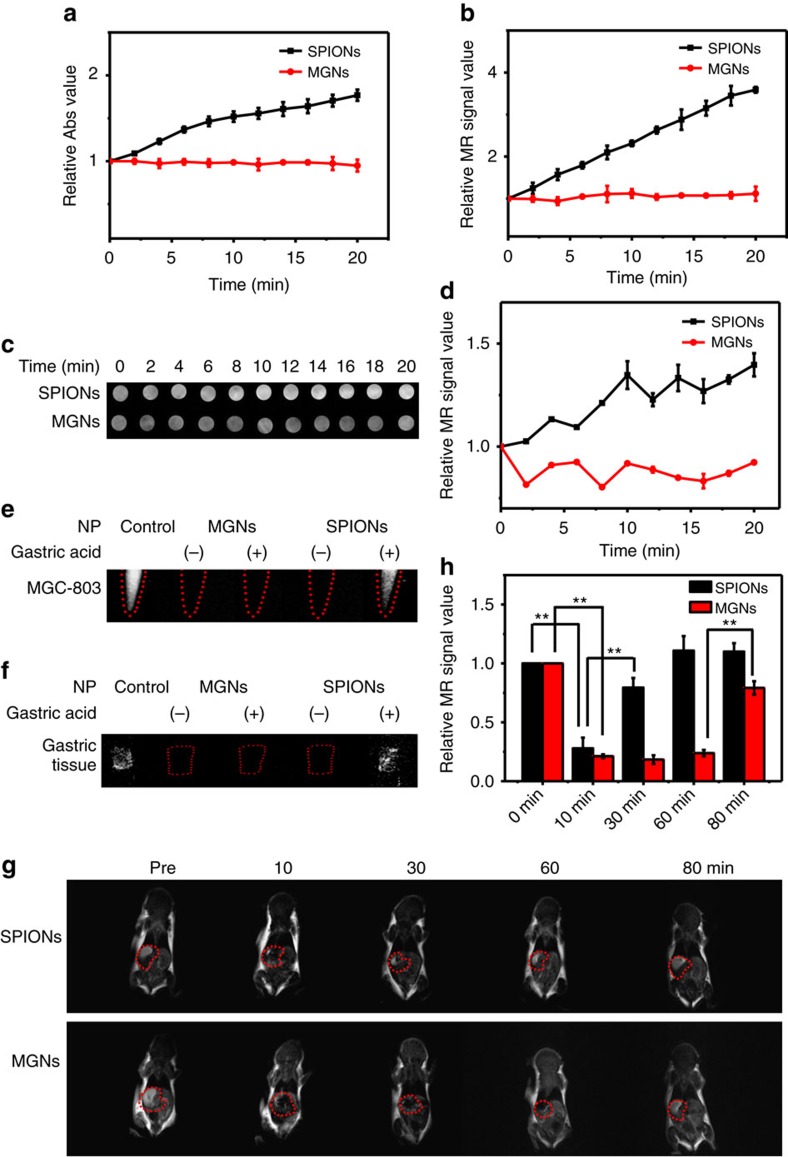
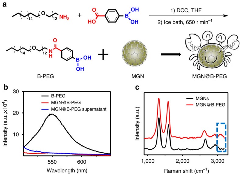
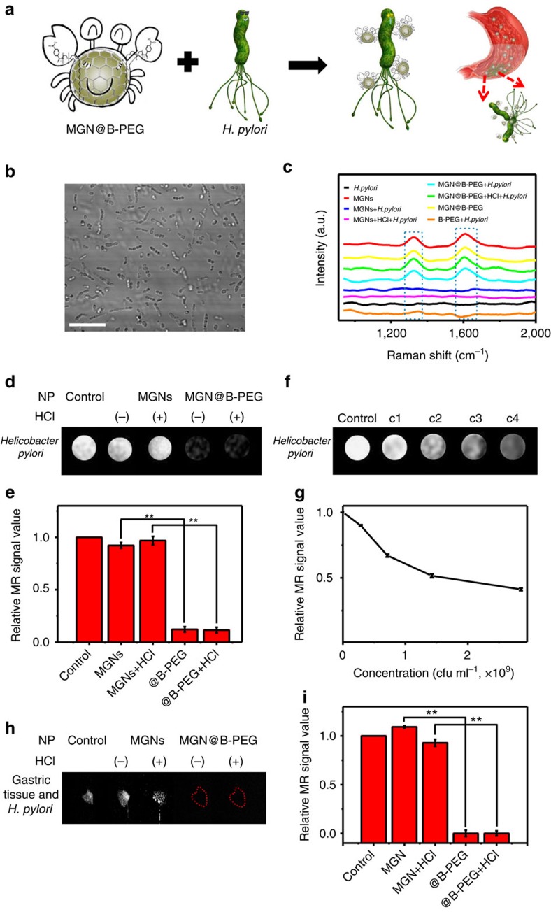
Helicobacter pylori infection is implicated in the aetiology of many diseases. Despite numerous studies, a painless, fast and direct method for the in situ detection of H. pylori remains a challenge, mainly due to the strong acidic/enzymatic environment of the gastric mucosa. Herein, we report the use of stable magnetic graphitic nanocapsules (MGNs), for in situ targeted magnetic resonance imaging (MRI) detection of H. pylori. Several layers of graphene as the shell effectively protect the magnetic core from corrosion while retaining the superior contrast effect for MRI in the gastric environment. Boronic-polyethylene glycol molecules were synthesized and modified on the MGN surface for targeted MRI detection. In a mouse model of H. pylori-induced infection, H. pylori was specifically detected through both T-weighted MR imaging and Raman gastric mucosa imaging using functionalized MGNs. These results indicated that enhancement of MRI using MGNs may be a promising diagnostic and bioimaging platform for very harsh conditions.
幽门螺杆菌感染与许多疾病的病因有关。尽管进行了大量研究,但由于胃黏膜的强酸/酶环境,仍然难以找到一种无痛、快速且直接的原位检测幽门螺杆菌的方法。在此,我们报告了使用稳定的磁性石墨纳米胶囊(MGN)进行原位靶向磁共振成像(MRI)检测幽门螺杆菌的方法。几层石墨烯作为外壳可以有效地保护磁核免受腐蚀,同时在胃环境中保持 MRI 的卓越对比效果。合成并修饰了硼-聚乙二醇分子在 MGN 表面上,用于靶向 MRI 检测。在幽门螺杆菌诱导感染的小鼠模型中,使用功能化的 MGN 通过 T 加权磁共振成像和拉曼胃黏膜成像特异性地检测到了幽门螺杆菌。这些结果表明,使用 MGN 增强 MRI 可能是一种有前途的诊断和生物成像平台,适用于非常恶劣的条件。