• 文献检索
  • 文档翻译
  • 深度研究
  • 学术资讯
  • Suppr Zotero 插件Zotero 插件
  • 邀请有礼
  • 套餐&价格
  • 历史记录
应用&插件
Suppr Zotero 插件Zotero 插件浏览器插件Mac 客户端Windows 客户端微信小程序
定价
高级版会员购买积分包购买API积分包
服务
文献检索文档翻译深度研究API 文档MCP 服务
关于我们
关于 Suppr公司介绍联系我们用户协议隐私条款
关注我们

Suppr 超能文献

核心技术专利:CN118964589B侵权必究
粤ICP备2023148730 号-1Suppr @ 2026

文献检索

告别复杂PubMed语法,用中文像聊天一样搜索,搜遍4000万医学文献。AI智能推荐,让科研检索更轻松。

立即免费搜索

文件翻译

保留排版,准确专业,支持PDF/Word/PPT等文件格式,支持 12+语言互译。

免费翻译文档

深度研究

AI帮你快速写综述,25分钟生成高质量综述,智能提取关键信息,辅助科研写作。

立即免费体验

人体胎盘血管树的整体器官血管铸型和 microCT 检查揭示了与妊娠疾病相关的新变化。

Whole organ vascular casting and microCT examination of the human placental vascular tree reveals novel alterations associated with pregnancy disease.

机构信息

Maternal and Fetal Health Research Centre, Faculty of Biology, Medicine and Health, University of Manchester, Manchester, United Kingdom.

Henry Moseley X-ray Imaging Facility, School of Materials, University of Manchester, Manchester, United Kingdom.

出版信息

Sci Rep. 2017 Jun 23;7(1):4144. doi: 10.1038/s41598-017-04379-0.

DOI:10.1038/s41598-017-04379-0
PMID:28646147
原文链接:https://pmc.ncbi.nlm.nih.gov/articles/PMC5482861/
Abstract

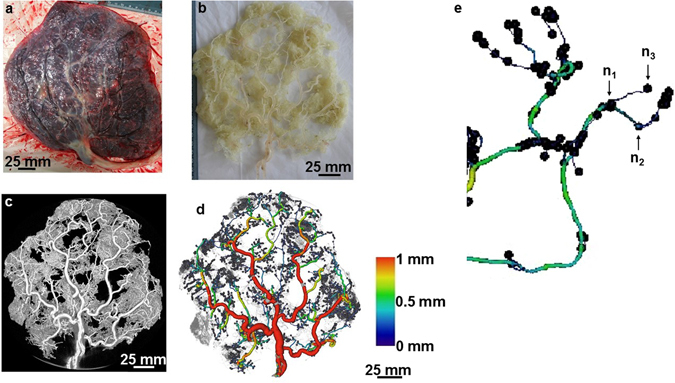
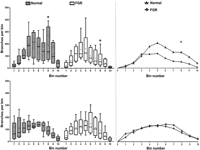
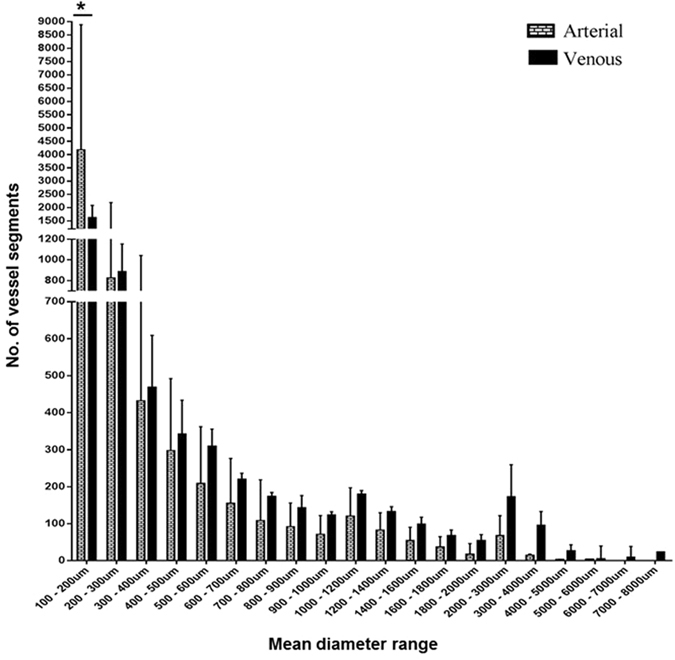
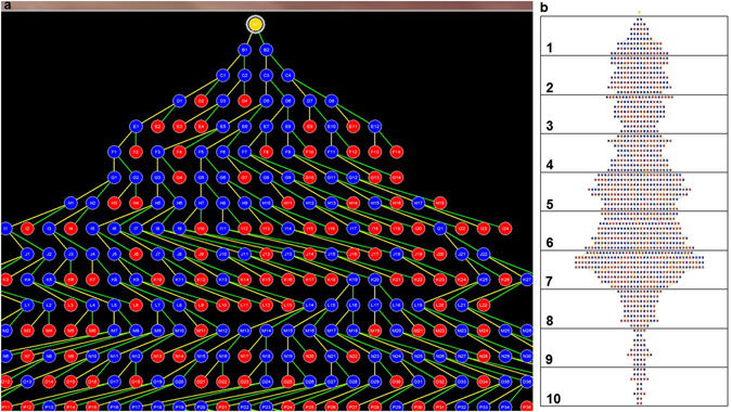
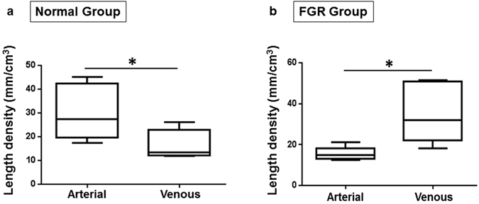
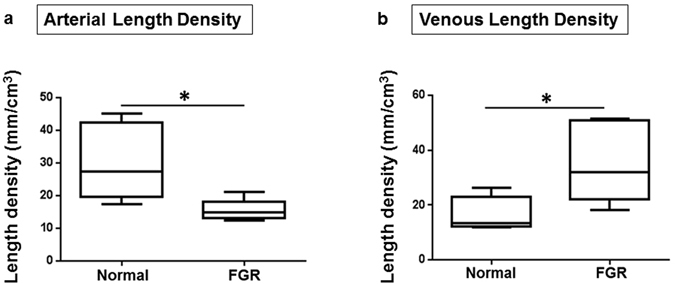
Experimental methods that allow examination of the intact vascular network of large organs, such as the human placenta are limited, preventing adequate comparison of normal and abnormal vascular development in pregnancy disease. Our aims were (i) to devise an effective technique for three-dimensional analyses of human placental vessels; (ii) demonstrate the utility of the technique in the comparison of placental vessel networks in normal and fetal growth restriction (FGR) complicated pregnancies. Radiopaque plastic vessel networks of normal and FGR placentas (n = 12/group) were created by filling the vessels with resin and corroding the surrounding tissues. Subsequently, each model was scanned in a microCT scanner, reconstructed into three-dimensional virtual objects and analysed in visualisation programmes. MicroCT imaging of the models defined vessel anatomy to our analyses threshold of 100 µm diameter. Median vessel length density was significantly shorter in arterial but longer in venous FGR networks compared to normals. No significant differences were demonstrable in arterial or venous tortuosity, diameter or branch density. This study demonstrates the potential effectiveness of microCT for ex-vivo examination of human placental vessel morphology. Our findings show significant discrepancies in vessel length density in FGR placentas. The effects on fetoplacental blood flow, and hence nutrient transfer to the fetus, are unknown.

摘要

实验方法限制了对大型器官(如人胎盘)完整血管网络的检查,从而无法充分比较妊娠疾病中正常和异常血管发育。我们的目的是:(i)设计一种有效的三维分析人胎盘血管的技术;(ii)展示该技术在比较正常和胎儿生长受限(FGR)复杂妊娠胎盘血管网络中的应用。通过用树脂填充血管并腐蚀周围组织,为正常和 FGR 胎盘制作了具有放射性不透明塑料血管网络的模型。随后,在 microCT 扫描仪中对每个模型进行扫描,在可视化程序中重建为三维虚拟对象并进行分析。模型的 microCT 成像将我们的分析阈值定义为 100µm 直径的血管解剖。与正常组相比,FGR 网络中的动脉血管长度密度明显缩短,而静脉血管长度密度则明显延长。在动脉或静脉的迂曲度、直径或分支密度方面,没有表现出显著差异。本研究证明了 microCT 对人胎盘血管形态进行离体检查的潜在有效性。我们的研究结果表明,FGR 胎盘的血管长度密度存在显著差异。但这对胎-胎盘血流的影响,以及对胎儿营养转移的影响尚不清楚。

https://cdn.ncbi.nlm.nih.gov/pmc/blobs/17a0/5482861/875ccd83ec94/41598_2017_4379_Fig6_HTML.jpg
https://cdn.ncbi.nlm.nih.gov/pmc/blobs/17a0/5482861/fff5aa512597/41598_2017_4379_Fig1_HTML.jpg
https://cdn.ncbi.nlm.nih.gov/pmc/blobs/17a0/5482861/c288bb83fddd/41598_2017_4379_Fig2_HTML.jpg
https://cdn.ncbi.nlm.nih.gov/pmc/blobs/17a0/5482861/265cd28f9a1b/41598_2017_4379_Fig3_HTML.jpg
https://cdn.ncbi.nlm.nih.gov/pmc/blobs/17a0/5482861/9523eb113f6e/41598_2017_4379_Fig4_HTML.jpg
https://cdn.ncbi.nlm.nih.gov/pmc/blobs/17a0/5482861/690a0185e841/41598_2017_4379_Fig5_HTML.jpg
https://cdn.ncbi.nlm.nih.gov/pmc/blobs/17a0/5482861/875ccd83ec94/41598_2017_4379_Fig6_HTML.jpg

相似文献

1
Whole organ vascular casting and microCT examination of the human placental vascular tree reveals novel alterations associated with pregnancy disease.人体胎盘血管树的整体器官血管铸型和 microCT 检查揭示了与妊娠疾病相关的新变化。
Sci Rep. 2017 Jun 23;7(1):4144. doi: 10.1038/s41598-017-04379-0.
2
Postpartum computed tomography angiography of the fetoplacental macrovasculature in normal pregnancies and in those complicated by fetal growth restriction.正常妊娠及胎儿生长受限孕妇胎盘胎儿大血管的产后 CT 血管造影
Acta Obstet Gynecol Scand. 2018 Mar;97(3):322-329. doi: 10.1111/aogs.13289.
3
Fetoplacental vascular alterations associated with fetal growth restriction.与胎儿生长受限相关的胎盘血管改变。
Placenta. 2014 Oct;35(10):808-15. doi: 10.1016/j.placenta.2014.07.013. Epub 2014 Aug 6.
4
Quantitative 3D micro-CT imaging of the human feto-placental vasculature in intrauterine growth restriction.宫内生长受限中人类胎儿-胎盘血管系统的定量三维显微CT成像
Placenta. 2008 Nov;29(11):937-41. doi: 10.1016/j.placenta.2008.08.017. Epub 2008 Oct 11.
5
Down-regulation of placental neuropilin-1 in fetal growth restriction.胎盘神经纤毛蛋白-1 在胎儿生长受限中的下调。
Am J Obstet Gynecol. 2016 Feb;214(2):279.e1-279.e9. doi: 10.1016/j.ajog.2015.09.068. Epub 2015 Sep 26.
6
Vascular network modeling reveals significant differences in vascular morphology in growth-restricted placentas.血管网络建模揭示了生长受限胎盘血管形态的显著差异。
Rev Obstet Gynecol. 2011;4(3-4):103-8.
7
Placental vascular sonobiopsy using three-dimensional power Doppler ultrasound in normal and growth restricted fetuses.在正常胎儿和生长受限胎儿中使用三维能量多普勒超声进行胎盘血管超声活检。
Placenta. 2009 May;30(5):391-7. doi: 10.1016/j.placenta.2009.02.010. Epub 2009 Mar 26.
8
Quantitative characterization of rodent feto-placental vasculature morphology in micro-computed tomography images.定量分析微计算机断层扫描图像中小鼠胎仔胎盘血管形态。
Comput Methods Programs Biomed. 2019 Oct;179:104984. doi: 10.1016/j.cmpb.2019.104984. Epub 2019 Jul 15.
9
Arterio-venous fetoplacental vascular geometry and hemodynamics in the mouse placenta.鼠胎盘的动静脉胎盘血管几何形状和血流动力学。
Placenta. 2017 Oct;58:46-51. doi: 10.1016/j.placenta.2017.08.007. Epub 2017 Aug 12.
10
Quantitative microCT imaging of a whole equine placenta and its blood vessel network.定量 microCT 成像技术对整个马胎盘及其血管网络的研究。
Placenta. 2024 Sep 2;154:216-219. doi: 10.1016/j.placenta.2024.07.313. Epub 2024 Jul 31.

引用本文的文献

1
Feto-placental blood vessel development.胎儿-胎盘血管发育
Development. 2025 Jun 1;152(11). doi: 10.1242/dev.204838. Epub 2025 Jun 2.
2
3D multiscale characterization of the human placenta: Bridging anatomy and histology by X-ray phase-contrast tomography.人胎盘的三维多尺度表征:通过X射线相衬断层扫描连接解剖学与组织学
PNAS Nexus. 2025 Jan 3;4(1):pgae583. doi: 10.1093/pnasnexus/pgae583. eCollection 2025 Jan.
3
μCT imaging of a multi-organ vascular fingerprint in rats.大鼠多器官血管指纹的 μCT 成像。

本文引用的文献

1
A multiscale model of placental oxygen exchange: The effect of villous tree structure on exchange efficiency.胎盘氧交换的多尺度模型:绒毛树结构对交换效率的影响。
J Theor Biol. 2016 Nov 7;408:1-12. doi: 10.1016/j.jtbi.2016.06.037. Epub 2016 Jul 1.
2
Novel 3D light microscopic analysis of IUGR placentas points to a morphological correlate of compensated ischemic placental disease in humans.对宫内生长受限胎盘进行的新型三维光学显微镜分析揭示了人类代偿性缺血性胎盘疾病的形态学关联。
Sci Rep. 2016 Apr 5;6:24004. doi: 10.1038/srep24004.
3
Does 2D-Histologic identification of villous types of human placentas at birth enable sensitive and reliable interpretation of 3D structure?
PLoS One. 2024 Oct 14;19(10):e0308601. doi: 10.1371/journal.pone.0308601. eCollection 2024.
4
A flexible generative algorithm for growing in silico placentas.一种灵活的生成算法,用于在计算机中生成胎盘。
PLoS Comput Biol. 2024 Oct 7;20(10):e1012470. doi: 10.1371/journal.pcbi.1012470. eCollection 2024 Oct.
5
Molecular regulators of defective placental and cardiovascular development in fetal growth restriction.胎儿生长受限中胎盘和心血管发育缺陷的分子调节因子。
Clin Sci (Lond). 2024 Jul 3;138(13):761-775. doi: 10.1042/CS20220428.
6
Blood flow and transport in the human placenta.人体胎盘的血流与物质运输
Annu Rev Fluid Mech. 2019 Jan;51:25-47. doi: 10.1146/annurev-fluid-010518-040219. Epub 2018 Aug 3.
7
Flow in fetoplacental-like microvessels in vitro enhances perfusion, barrier function, and matrix stability.体外胎盘中类似微血管的血流增强了灌注、屏障功能和基质稳定性。
Sci Adv. 2023 Dec 22;9(51):eadj8540. doi: 10.1126/sciadv.adj8540.
8
Human placental vascular and perivascular cell heterogeneity differs between first trimester and term, and in pregnancies affected by foetal growth restriction.人类胎盘血管和血管周围细胞的异质性在早孕期和足月时不同,并且在受胎儿生长受限影响的妊娠中也不同。
Mol Hum Reprod. 2023 Nov 29;29(12). doi: 10.1093/molehr/gaad041.
9
Fetal-placental MR angiography at 1.5 T and 3 T.1.5T 和 3T 下胎儿-胎盘磁共振血管成像。
Magn Reson Imaging. 2023 Oct;102:133-140. doi: 10.1016/j.mri.2023.05.003. Epub 2023 May 18.
10
Magnetic Resonance Imaging Angiography of Physiological and Pathological Pregnancy Placentas Ex Vivo: Protocol for a Prospective Pilot Study.生理性和病理性妊娠胎盘离体磁共振成像血管造影:一项前瞻性试点研究方案
JMIR Res Protoc. 2022 Aug 10;11(8):e35051. doi: 10.2196/35051.
出生时对人胎盘绒毛类型进行二维组织学鉴定能否实现对三维结构的灵敏且可靠的解读?
Placenta. 2015 Dec;36(12):1425-32. doi: 10.1016/j.placenta.2015.10.003. Epub 2015 Oct 19.
4
Classification of placental lesions.胎盘病变的分类。
Am J Obstet Gynecol. 2015 Oct;213(4 Suppl):S21-8. doi: 10.1016/j.ajog.2015.05.056.
5
Dysregulated flow-mediated vasodilatation in the human placenta in fetal growth restriction.胎儿生长受限中人类胎盘血流介导的血管舒张功能失调。
J Physiol. 2015 Jul 15;593(14):3077-92. doi: 10.1113/JP270495. Epub 2015 Jun 8.
6
Intrauterine growth restriction is associated with structural alterations in human umbilical cord and decreased nitric oxide-induced relaxation of umbilical vein.宫内生长受限与人类脐带的结构改变以及一氧化氮诱导的脐静脉舒张功能降低有关。
Placenta. 2014 Nov;35(11):891-9. doi: 10.1016/j.placenta.2014.08.090. Epub 2014 Sep 2.
7
Novel 3D microscopic analysis of human placental villous trees reveals unexpected significance of branching angles.对人胎盘绒毛树的新型三维显微镜分析揭示了分支角度出人意料的重要性。
Sci Rep. 2014 Aug 26;4:6192. doi: 10.1038/srep06192.
8
Fetoplacental vascular alterations associated with fetal growth restriction.与胎儿生长受限相关的胎盘血管改变。
Placenta. 2014 Oct;35(10):808-15. doi: 10.1016/j.placenta.2014.07.013. Epub 2014 Aug 6.
9
Sonographic measurement of the umbilical cord area and the diameters of its vessels during pregnancy.孕期超声测量脐带面积及其血管直径。
J Obstet Gynaecol. 2012 Apr;32(3):230-6. doi: 10.3109/01443615.2011.647129.
10
A method for quantitative characterization of growth in the 3-D structure of rat pulmonary arteries.一种定量描述大鼠肺内动脉 3D 结构生长的方法。
Microvasc Res. 2012 Mar;83(2):146-53. doi: 10.1016/j.mvr.2011.12.003. Epub 2012 Jan 2.