Wang Hesheng, Chandarana Hersh, Block Kai Tobias, Vahle Thomas, Fenchel Matthias, Das Indra J
Department of Radiation Oncology, New York University School of Medicine, Langone Medical Center, New York, NY, USA.
Bernard and Irene Schwartz Center for Biomedical Imaging, Department of Radiology, New York University School of Medicine, New York, NY, USA.
Radiat Oncol. 2017 Jun 26;12(1):108. doi: 10.1186/s13014-017-0845-5.
Interest in MR-only treatment planning for radiation therapy is growing rapidly with the emergence of integrated MRI/linear accelerator technology. The purpose of this study was to evaluate the feasibility of using synthetic CT images generated from conventional Dixon-based MRI scans for radiation treatment planning of lung cancer.
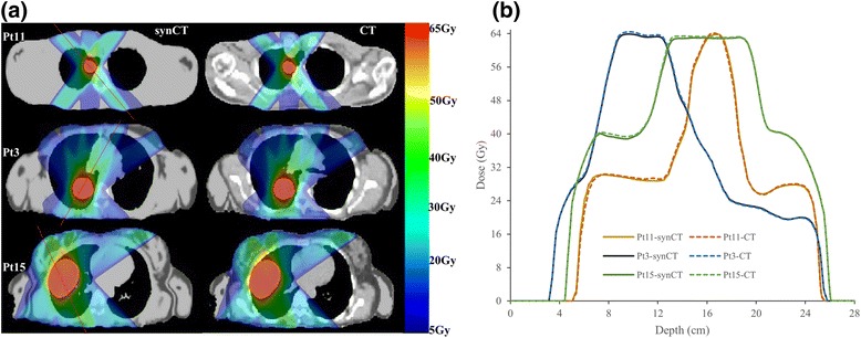
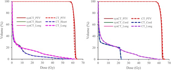
Eleven patients who underwent whole-body PET/MR imaging following a PET/CT exam were randomly selected from an ongoing prospective IRB-approved study. Attenuation maps derived from the Dixon MR Images and atlas-based method was used to create CT data (synCT). Treatment planning for radiation treatment of lung cancer was optimized on the synCT and subsequently copied to the registered CT (planCT) for dose calculation. Planning target volumes (PTVs) with three sizes and four different locations in the lung were planned for irradiation. The dose-volume metrics comparison and 3D gamma analysis were performed to assess agreement between the synCT and CT calculated dose distributions.
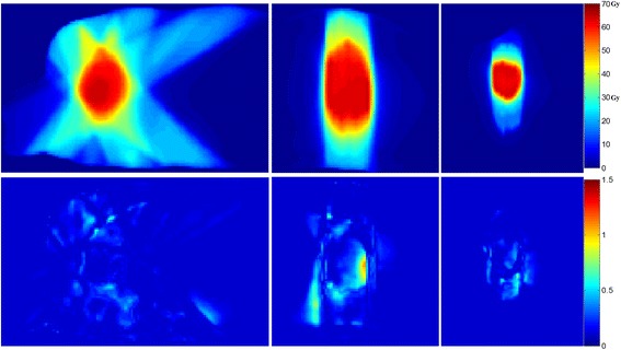
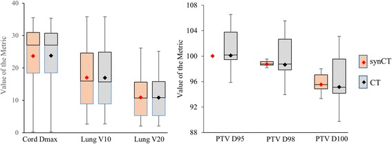
Mean differences between PTV doses on synCT and CT across all the plans were -0.1% ± 0.4%, 0.1% ± 0.5%, and 0.4% ± 0.5% for D95, D98 and D100, respectively. Difference in dose between the two datasets for organs at risk (OARs) had average differences of -0.14 ± 0.07 Gy, 0.0% ± 0.1%, and -0.1% ± 0.2% for maximum spinal cord, lung V20, and heart V40 respectively. In patient groups based on tumor size and location, no significant differences were observed in the PTV and OARs dose-volume metrics (p > 0.05), except for the maximum spinal-cord dose when the target volumes were located at the lung apex (p = 0.001). Gamma analysis revealed a pass rate of 99.3% ± 1.1% for 2%/2 mm (dose difference/distance to agreement) acceptance criteria in every plan.
The synCT generated from Dixon-based MRI allows for dose calculation of comparable accuracy to the standard CT for lung cancer treatment planning. The dosimetric agreement between synCT and CT calculated doses warrants further development of a MR-only workflow for radiotherapy of lung cancer.
随着集成式MRI/直线加速器技术的出现,仅基于磁共振成像(MR)的放射治疗计划的关注度正在迅速增长。本研究的目的是评估使用基于传统狄克逊(Dixon)法MRI扫描生成的合成CT图像进行肺癌放射治疗计划的可行性。
从一项正在进行的、经机构审查委员会(IRB)批准的前瞻性研究中,随机选取11例在PET/CT检查后接受全身PET/MR成像的患者。采用从狄克逊MR图像和基于图谱的方法得出的衰减图来创建CT数据(合成CT)。在合成CT上对肺癌放射治疗计划进行优化,随后将其复制到已配准的CT(计划CT)上进行剂量计算。针对肺内三种大小和四个不同位置的计划靶区(PTV)制定照射计划。进行剂量体积指标比较和三维伽马分析,以评估合成CT和CT计算的剂量分布之间的一致性。
在所有计划中,合成CT和CT上PTV剂量的平均差异,对于D95、D98和D100分别为-0.1%±0.4%、0.1%±0.5%和0.4%±0.5%。两个数据集之间危及器官(OAR)剂量的差异,对于脊髓最大剂量、肺V20和心脏V40,平均差异分别为-0.14±0.07 Gy、0.0%±0.1%和-0.1%±0.2%。在基于肿瘤大小和位置的患者组中,PTV和OAR剂量体积指标未观察到显著差异(p>0.05),但当靶区位于肺尖时,脊髓最大剂量除外(p = 0.001)。伽马分析显示,在每个计划中,对于2%/2 mm(剂量差异/一致性距离)的接受标准,通过率为99.3%±1.1%。
基于狄克逊法MRI生成的合成CT在肺癌治疗计划中能够实现与标准CT相当的剂量计算精度。合成CT和CT计算剂量之间的剂量学一致性为肺癌放射治疗仅基于MR的工作流程的进一步发展提供了依据。