Shen Xue, Li Tingting, Chen Zhongyuan, Geng Yue, Xie Xiaoxue, Li Shun, Yang Hong, Wu Chunhui, Liu Yiyao
Department of Biophysics, School of Life Science and Technology.
Center for Information in Biology, University of Electronic Science and Technology of China, Chengdu, Sichuan, People's Republic of China.
Int J Nanomedicine. 2017 Jun 6;12:4299-4322. doi: 10.2147/IJN.S136766. eCollection 2017.
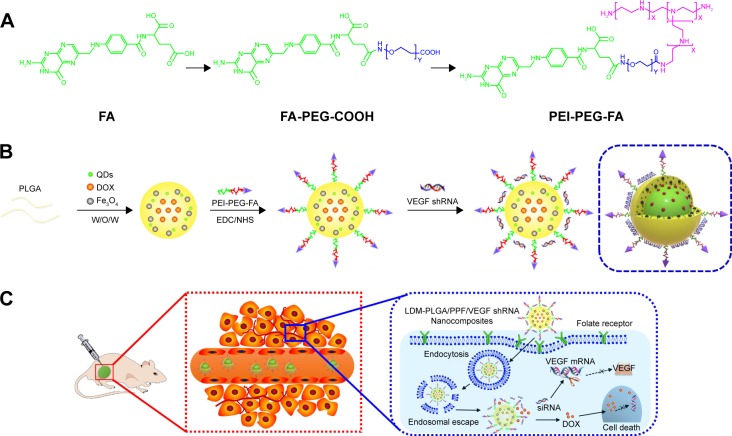
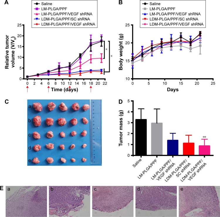
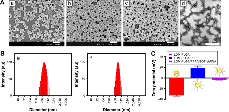
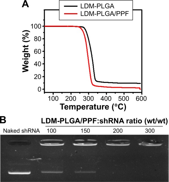
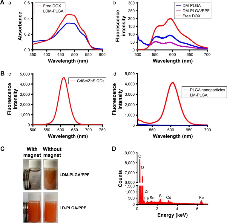
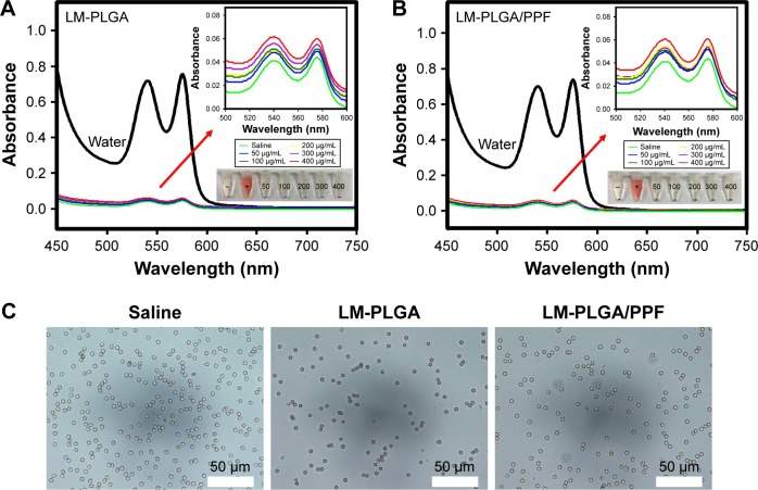
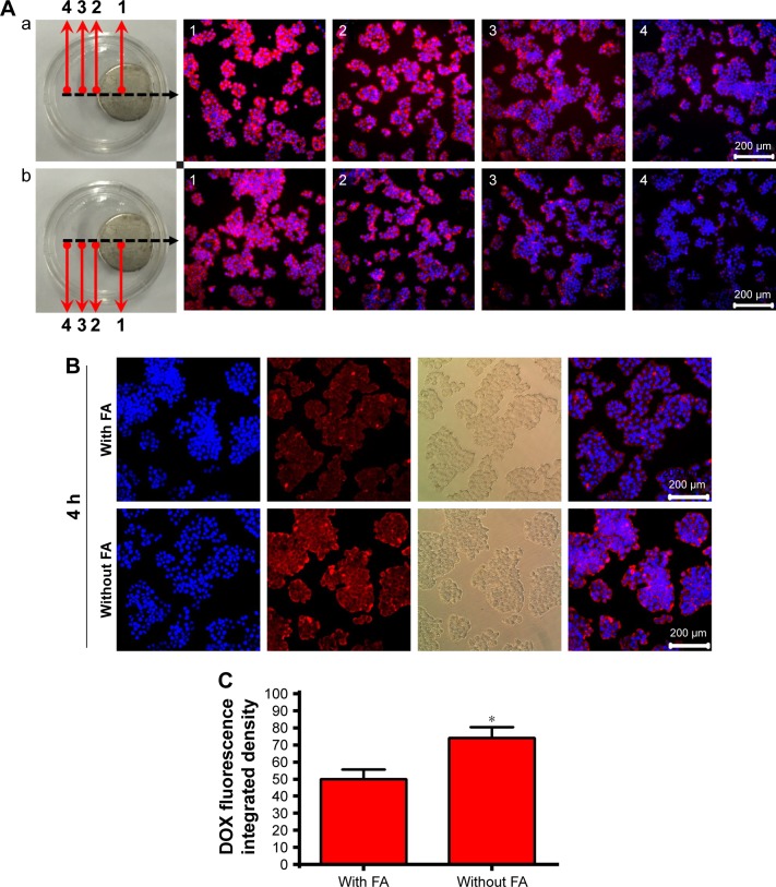
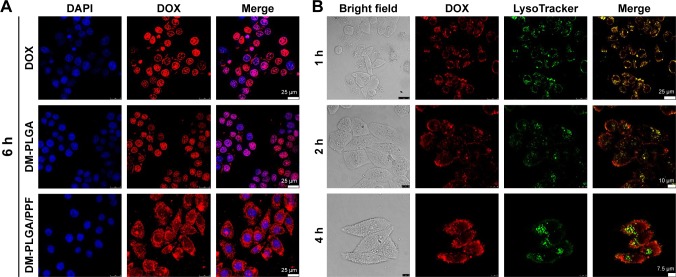
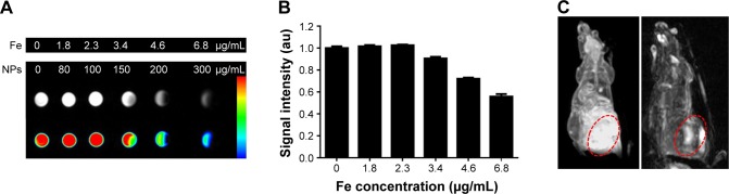
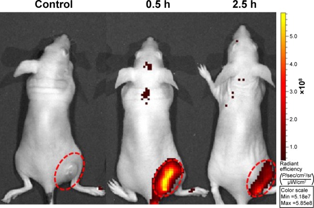
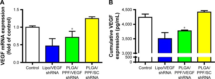
Cancer diagnosis and treatment represent an urgent medical need given the rising cancer incidence over the past few decades. Cancer theranostics, namely, the combination of diagnostics and therapeutics within a single agent, are being developed using various anticancer drug-, siRNA-, or inorganic materials-loaded nanocarriers. Herein, we demonstrate a strategy of encapsulating quantum dots, superparamagnetic FeO nanocrystals, and doxorubicin (DOX) into biodegradable poly(d,l-lactic--glycolic acid) (PLGA) polymeric nanocomposites using the double emulsion solvent evaporation method, followed by coupling to the amine group of polyethyleneimine premodified with polyethylene glycol-folic acid (PEI-PEG-FA [PPF]) segments and adsorption of vascular endothelial growth factor (VEGF)-targeted small hairpin RNA (shRNA). VEGF is important for tumor growth, progression, and metastasis. These drug-loaded luminescent/magnetic PLGA-based hybrid nanocomposites (LDM-PLGA/PPF/VEGF shRNA) were fabricated for tumor-specific targeting, drug/gene delivery, and cancer imaging. The data showed that LDM-PLGA/PPF/VEGF shRNA nanocomposites can codeliver DOX and VEGF shRNA into tumor cells and effectively suppress VEGF expression, exhibiting remarkable synergistic antitumor effects both in vitro and in vivo. The cell viability waŝ14% when treated with LDM-PLGA/PPF/VEGF shRNA nanocomposites ([DOX] =25 μg/mL), and in vivo tumor growth data showed that the tumor volume decreased by 81% compared with the saline group at 21 days postinjection. Magnetic resonance and fluorescence imaging data revealed that the luminescent/magnetic hybrid nanocomposites may also be used as an efficient nanoprobe for enhanced -weighted magnetic resonance and fluorescence imaging in vitro and in vivo. The present work validates the great potential of the developed multifunctional LDM-PLGA/PPF/VEGF shRNA nanocomposites as effective theranostic agents through the codelivery of drugs/genes and dual-modality imaging in cancer treatment.
鉴于在过去几十年中癌症发病率不断上升,癌症的诊断和治疗成为了一项迫切的医疗需求。癌症诊疗一体化,即在单一制剂中结合诊断和治疗功能,正通过各种负载抗癌药物、小干扰RNA(siRNA)或无机材料的纳米载体进行开发。在此,我们展示了一种策略,即使用双乳液溶剂蒸发法将量子点、超顺磁性FeO纳米晶体和阿霉素(DOX)封装到可生物降解的聚(d,l-乳酸-乙醇酸)(PLGA)聚合物纳米复合材料中,随后与用聚乙二醇-叶酸(PEI-PEG-FA [PPF])片段预修饰的聚乙烯亚胺的胺基偶联,并吸附血管内皮生长因子(VEGF)靶向的小发夹RNA(shRNA)。VEGF对肿瘤的生长、进展和转移至关重要。这些负载药物的基于PLGA的发光/磁性杂化纳米复合材料(LDM-PLGA/PPF/VEGF shRNA)被制备用于肿瘤特异性靶向、药物/基因递送和癌症成像。数据表明,LDM-PLGA/PPF/VEGF shRNA纳米复合材料可以将DOX和VEGF shRNA共递送至肿瘤细胞,并有效抑制VEGF表达,在体外和体内均表现出显著的协同抗肿瘤作用。用LDM-PLGA/PPF/VEGF shRNA纳米复合材料([DOX]=25μg/mL)处理时,细胞活力为14%,体内肿瘤生长数据显示,注射后21天时,肿瘤体积与盐水组相比减少了81%。磁共振和荧光成像数据表明,发光/磁性杂化纳米复合材料也可用作一种有效的纳米探针,用于体外和体内的加权磁共振增强和荧光成像。目前的工作通过在癌症治疗中药物/基因的共递送和双模态成像,验证了所开发的多功能LDM-PLGA/PPF/VEGF shRNA纳米复合材料作为有效诊疗试剂的巨大潜力。