Department of Cardiology, The First Affiliated Hospital of South China University, Hengyang, Hunan 421001, P.R. China.
Department of Pharmacy, The Second Affiliated Hospital of South China University, Hengyang, Hunan 421001, P.R. China.
Mol Med Rep. 2017 Aug;16(2):1715-1722. doi: 10.3892/mmr.2017.6813. Epub 2017 Jun 20.
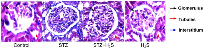
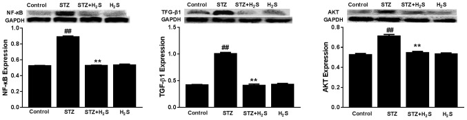
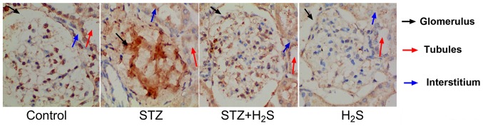
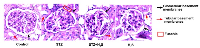
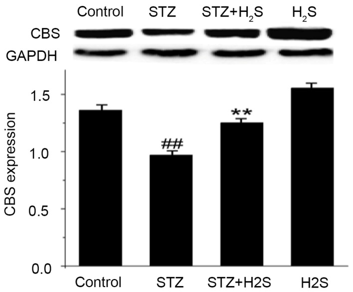
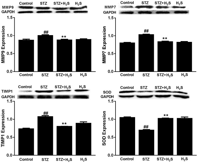
The present study aimed to explore the effect of hydrogen sulfide (H2S) on renal tissue fibrosis and its mechanism in diabetic rats. Rats were randomly divided into four groups (n=13/group): Control group; induced diabetes mellitus group (STZ); induced diabetes mellitus treated with H2S group (STZ + H2S); normal rats treated with H2S group (H2S). The diabetic model was induced by intraperitoneal (i.p.) injections of 40 mg/kg body weight streptozotocin (STZ); the control group was treated with saline every day (i.p); NaHS (100 µmol/kg i.p.) was administered to rats of STZ + H2S group and H2S group. After 8 weeks, rat body weight and 24 h proteinuria levels were determined in each group, renal pathological morphology was analyzed by Masson's trichrome staining, collagen IV content was detected by immunohistochemistry, and periodic acid‑Schiff (PAS) staining was performed on renal glomerular and tubular basement membranes. The expression levels of matrix metalloproteinase 9 (MMP9), MMP7, tissue inhibitor of metalloproteinase 1 (TIMP1), superoxide dismutase (SOD), serine/threonine kinase AKT, transforming growth factor (TGF)‑β1, nuclear factor (NF)‑κB and several autophagy related proteins were assessed by western blot analysis. Compared with the control group, renal tissue fibrosis was observed, collagen IV expression and the 24 h proteinuria quantity was markedly increased and the amount of PAS positive material in renal glomerular and tubular basement membranes was notably increased in STZ‑treated rats. Furthermore, the expression levels of MMP9, MMP7, TIMP1, autophagy‑associated proteins, AKT, TGF‑β1 and NF‑κB protein were significantly increased, and SOD expression levels were significantly decreased in the STZ group compared with the control (P<0.05). In the H2S+STZ group, renal tissue fibrosis and the expression of collagen IV were improved, 24 h proteinuria was decreased, the amount of PAS positive material in renal glomerular and tubular basement membranes was decreased, the expression levels MMP9, MMP7, TIMP1, autophagy‑associated proteins, AKT, TGF‑β1 and NF‑κB protein were significantly decreased, and the expression levels of SOD were significantly increased compared with the STZ group (P<0.05). In conclusion, H2S may improve renal tissue fibrosis by inhibiting autophagy, upregulating SOD and downregulating AKT, TGF‑β1 and NF-κB.
本研究旨在探讨硫化氢(H2S)对糖尿病大鼠肾组织纤维化的影响及其机制。大鼠随机分为四组(每组 13 只):对照组;诱导型糖尿病组(STZ);STZ 诱导型糖尿病治疗组(STZ+H2S);正常大鼠 H2S 治疗组(H2S)。采用腹腔内(i.p.)注射 40mg/kg 体重链脲佐菌素(STZ)诱导糖尿病模型;对照组每天给予生理盐水(i.p.);STZ+H2S 组和 H2S 组大鼠给予 NaHS(100μmol/kg i.p.)。8 周后,测定各组大鼠体重和 24h 尿蛋白水平,采用 Masson 三色染色法分析肾组织病理形态,免疫组织化学法检测胶原 IV 含量,过碘酸雪夫(PAS)染色法检测肾肾小球和肾小管基底膜 PAS 阳性物质。采用 Western blot 分析检测基质金属蛋白酶 9(MMP9)、MMP7、金属蛋白酶组织抑制剂 1(TIMP1)、超氧化物歧化酶(SOD)、丝氨酸/苏氨酸激酶 AKT、转化生长因子(TGF)-β1、核因子(NF)-κB 和几种自噬相关蛋白的表达水平。与对照组相比,STZ 处理大鼠的肾组织纤维化、胶原 IV 表达和 24h 尿蛋白量明显增加,肾肾小球和肾小管基底膜 PAS 阳性物质明显增加。此外,与对照组相比,STZ 组 MMP9、MMP7、TIMP1、自噬相关蛋白、AKT、TGF-β1 和 NF-κB 蛋白表达水平显著升高,SOD 表达水平显著降低(P<0.05)。在 H2S+STZ 组,肾组织纤维化和胶原 IV 表达得到改善,24h 尿蛋白减少,肾肾小球和肾小管基底膜 PAS 阳性物质减少,MMP9、MMP7、TIMP1、自噬相关蛋白、AKT、TGF-β1 和 NF-κB 蛋白表达水平显著降低,SOD 表达水平显著升高与 STZ 组相比(P<0.05)。综上所述,H2S 可能通过抑制自噬、上调 SOD 和下调 AKT、TGF-β1 和 NF-κB 来改善肾组织纤维化。