• 文献检索
  • 文档翻译
  • 深度研究
  • 学术资讯
  • Suppr Zotero 插件Zotero 插件
  • 邀请有礼
  • 套餐&价格
  • 历史记录
应用&插件
Suppr Zotero 插件Zotero 插件浏览器插件Mac 客户端Windows 客户端微信小程序
定价
高级版会员购买积分包购买API积分包
服务
文献检索文档翻译深度研究API 文档MCP 服务
关于我们
关于 Suppr公司介绍联系我们用户协议隐私条款
关注我们

Suppr 超能文献

核心技术专利:CN118964589B侵权必究
粤ICP备2023148730 号-1Suppr @ 2026

文献检索

告别复杂PubMed语法,用中文像聊天一样搜索,搜遍4000万医学文献。AI智能推荐,让科研检索更轻松。

立即免费搜索

文件翻译

保留排版,准确专业,支持PDF/Word/PPT等文件格式,支持 12+语言互译。

免费翻译文档

深度研究

AI帮你快速写综述,25分钟生成高质量综述,智能提取关键信息,辅助科研写作。

立即免费体验

吉西他滨功能化磁铁矿纳米颗粒的制备及其细胞毒性

Fabrication and Cytotoxicity of Gemcitabine-Functionalized Magnetite Nanoparticles.

作者信息

Popescu Roxana Cristina, Andronescu Ecaterina, Vasile Bogdan Ștefan, Truşcă Roxana, Boldeiu Adina, Mogoantă Laurențiu, Mogoșanu George Dan, Temelie Mihaela, Radu Mihai, Grumezescu Alexandru Mihai, Savu Diana

机构信息

Department of Life and Environmental Physics, "Horia Hulubei" National Institute for Physics and Nuclear Engineering, 30 Reactorului Street, Măgurele 077125, Romania.

Department of Science and Engineering of Oxide Materials and Nanomaterials, Faculty of Applied Chemistry and Materials Science, University Politehnica of Bucharest, 1-7 Polizu Street, Bucharest 011061, Romania.

出版信息

Molecules. 2017 Jun 28;22(7):1080. doi: 10.3390/molecules22071080.

DOI:10.3390/molecules22071080
PMID:28657606
原文链接:https://pmc.ncbi.nlm.nih.gov/articles/PMC6152359/
Abstract

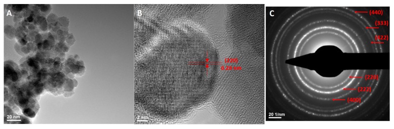
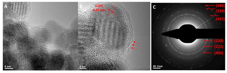
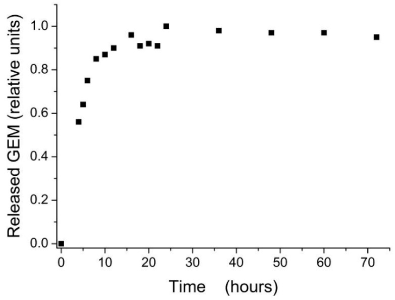
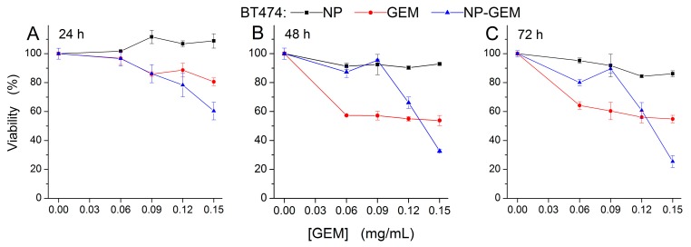
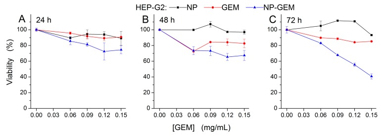
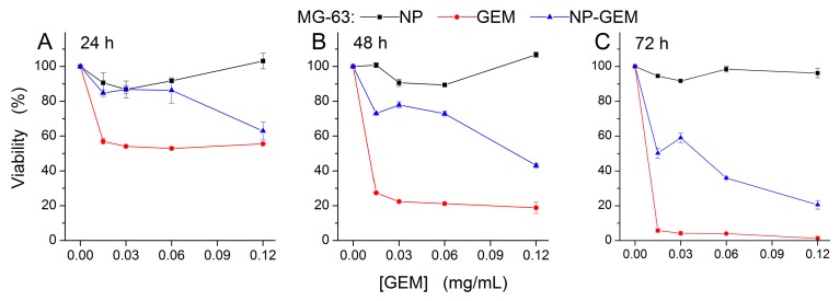
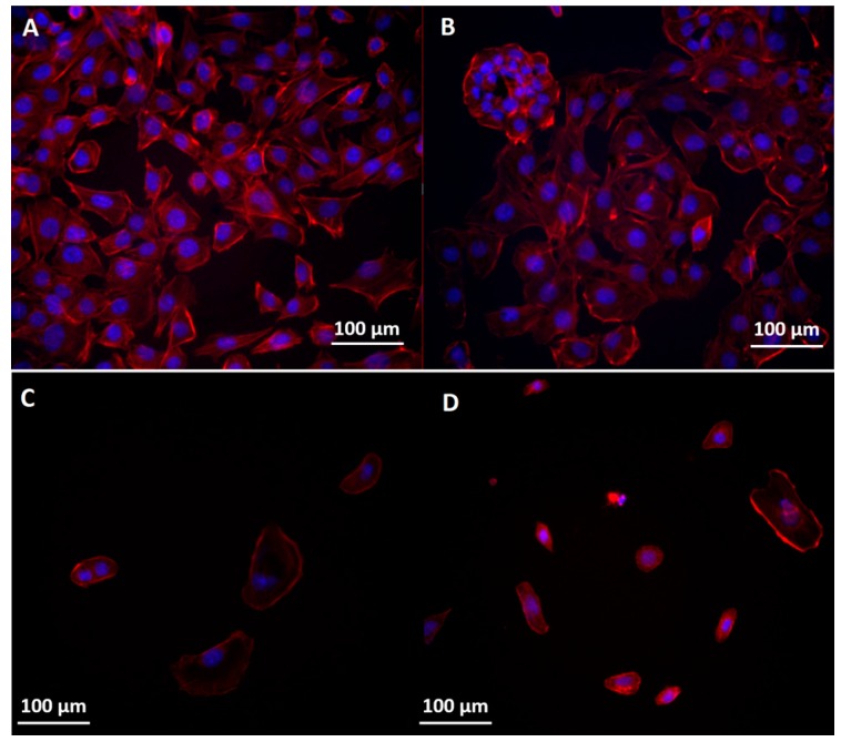
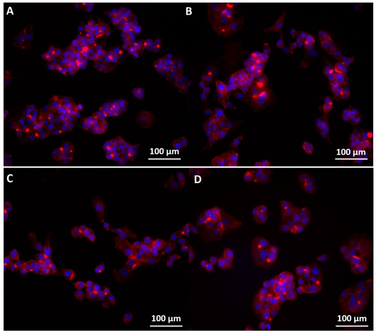
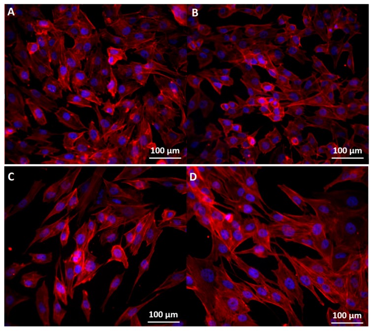
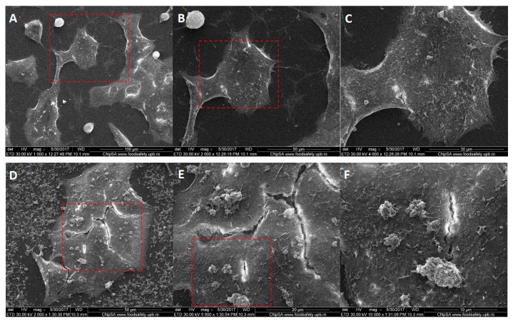
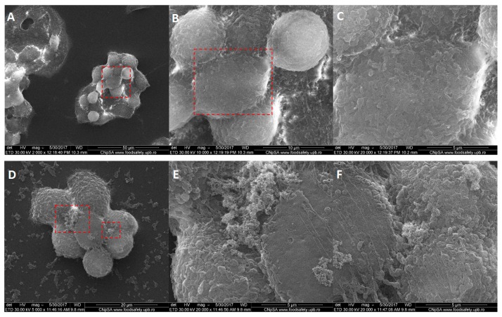
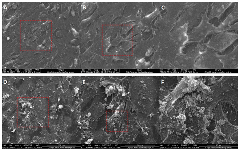
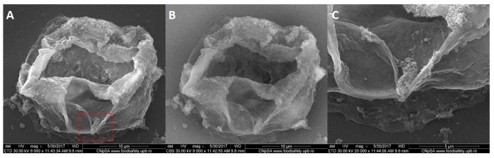
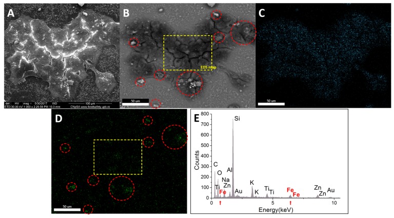
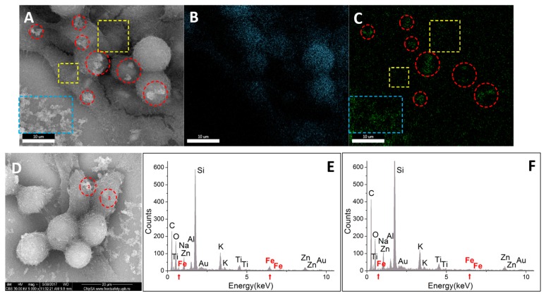
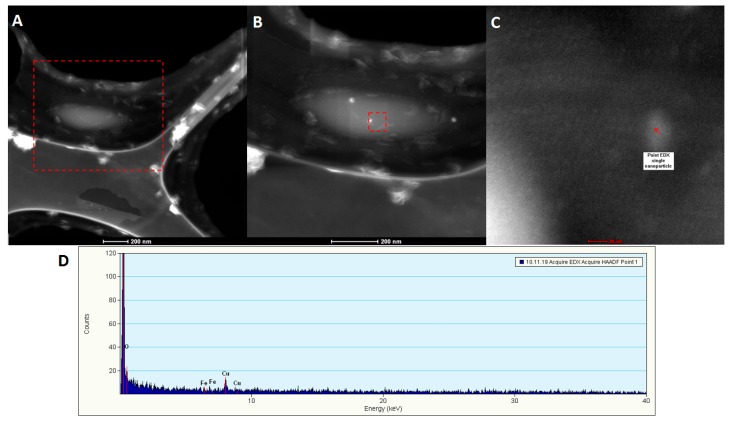
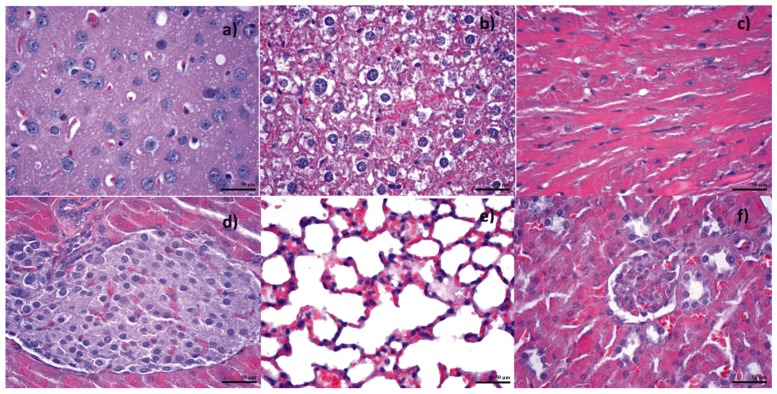
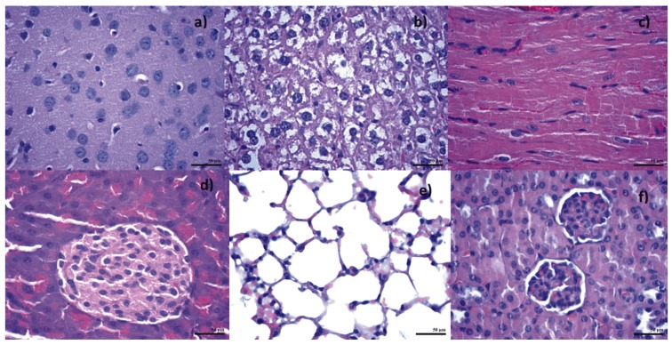
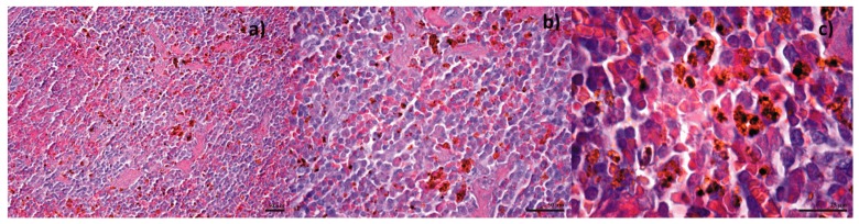
Nanotechnology has been successfully used for the fabrication of targeted anti-cancer drug carriers. This study aimed to obtain Fe₃O₄ nanoparticles functionalized with Gemcitabine to improve the cytotoxic effects of the chemotherapeutic substance on cancer cells. The (un) functionalized magnetite nanoparticles were synthesized using a modified co-precipitation method. The nanoconjugate characterization was performed by XRD, SEM, SAED and HRTEM; the functionalizing of magnetite with anti-tumor substances has been highlighted through TGA. The interaction with biologic media has been studied by means of stability and agglomeration tendency (using DLS and Zeta Potential); also, the release kinetics of the drug in culture media was evaluated. Cytotoxicity of free-Gemcitabine and the obtained nanoconjugate were evaluated on human BT 474 breast ductal carcinoma, HepG2 hepatocellular carcinoma and MG 63 osteosarcoma cells by MTS. In parallel, cellular morphology of these cells were examined through fluorescence microscopy and SEM. The localization of the nanoparticles related to the cells was studied using SEM, EDX and TEM. Hemolysis assay showed no damage of erythrocytes. Additionally, an in vivo biodistribution study was made for tracking where Fe₃O₄@Gemcitabine traveled in the body of mice. Our results showed that the transport of the drug improves the cytotoxic effects in comparison with the one produced by free Gemcitabine for the BT474 and HepG2 cells. The in vivo biodistribution test proved nanoparticle accumulation in the vital organs, with the exception of spleen, where black-brown deposits have been found. These results indicate that our Gemcitabine-functionalized nanoparticles are a promising targeted system for applications in cancer therapy.

摘要

纳米技术已成功用于制备靶向抗癌药物载体。本研究旨在获得用吉西他滨功能化的Fe₃O₄纳米颗粒,以提高这种化疗物质对癌细胞的细胞毒性作用。采用改进的共沉淀法合成了(未)功能化的磁铁矿纳米颗粒。通过XRD、SEM、SAED和HRTEM对纳米共轭物进行了表征;通过TGA突出了用抗肿瘤物质对磁铁矿进行功能化的过程。通过稳定性和团聚趋势(使用DLS和Zeta电位)研究了与生物介质的相互作用;此外,还评估了药物在培养基中的释放动力学。通过MTS评估了游离吉西他滨和所得纳米共轭物对人BT 474乳腺导管癌、HepG2肝细胞癌和MG 63骨肉瘤细胞的细胞毒性。同时,通过荧光显微镜和SEM检查了这些细胞的细胞形态。使用SEM、EDX和TEM研究了纳米颗粒与细胞的定位关系。溶血试验表明红细胞未受损。此外,还进行了体内生物分布研究,以追踪Fe₃O₄@吉西他滨在小鼠体内的行踪。我们的结果表明,与游离吉西他滨对BT474和HepG2细胞产生的作用相比,药物的转运提高了细胞毒性作用。体内生物分布试验证明纳米颗粒在重要器官中积累,但脾脏除外,在脾脏中发现了黑褐色沉积物。这些结果表明,我们的吉西他滨功能化纳米颗粒是一种有前途的靶向系统,可用于癌症治疗。

https://cdn.ncbi.nlm.nih.gov/pmc/blobs/f747/6152359/5767cb940072/molecules-22-01080-g024.jpg
https://cdn.ncbi.nlm.nih.gov/pmc/blobs/f747/6152359/0b1430f14495/molecules-22-01080-g001.jpg
https://cdn.ncbi.nlm.nih.gov/pmc/blobs/f747/6152359/89616fb07486/molecules-22-01080-g002.jpg
https://cdn.ncbi.nlm.nih.gov/pmc/blobs/f747/6152359/1306cd042819/molecules-22-01080-g003.jpg
https://cdn.ncbi.nlm.nih.gov/pmc/blobs/f747/6152359/4da066e75daa/molecules-22-01080-g004.jpg
https://cdn.ncbi.nlm.nih.gov/pmc/blobs/f747/6152359/7f2c82c22aaa/molecules-22-01080-g005.jpg
https://cdn.ncbi.nlm.nih.gov/pmc/blobs/f747/6152359/bdd578463a01/molecules-22-01080-g006.jpg
https://cdn.ncbi.nlm.nih.gov/pmc/blobs/f747/6152359/48391ec60411/molecules-22-01080-g007.jpg
https://cdn.ncbi.nlm.nih.gov/pmc/blobs/f747/6152359/e27951f6a652/molecules-22-01080-g008.jpg
https://cdn.ncbi.nlm.nih.gov/pmc/blobs/f747/6152359/58590a80142d/molecules-22-01080-g009.jpg
https://cdn.ncbi.nlm.nih.gov/pmc/blobs/f747/6152359/eb1795cef88d/molecules-22-01080-g010.jpg
https://cdn.ncbi.nlm.nih.gov/pmc/blobs/f747/6152359/1062a737f1c9/molecules-22-01080-g011.jpg
https://cdn.ncbi.nlm.nih.gov/pmc/blobs/f747/6152359/aadf8c649c1f/molecules-22-01080-g012.jpg
https://cdn.ncbi.nlm.nih.gov/pmc/blobs/f747/6152359/825e84450fa6/molecules-22-01080-g013.jpg
https://cdn.ncbi.nlm.nih.gov/pmc/blobs/f747/6152359/7ee5a66746e4/molecules-22-01080-g014.jpg
https://cdn.ncbi.nlm.nih.gov/pmc/blobs/f747/6152359/289a183ac973/molecules-22-01080-g015.jpg
https://cdn.ncbi.nlm.nih.gov/pmc/blobs/f747/6152359/084ab7187349/molecules-22-01080-g016.jpg
https://cdn.ncbi.nlm.nih.gov/pmc/blobs/f747/6152359/ed2a92651bbc/molecules-22-01080-g017.jpg
https://cdn.ncbi.nlm.nih.gov/pmc/blobs/f747/6152359/03473ba9a067/molecules-22-01080-g018.jpg
https://cdn.ncbi.nlm.nih.gov/pmc/blobs/f747/6152359/aef5cacd9c63/molecules-22-01080-g019.jpg
https://cdn.ncbi.nlm.nih.gov/pmc/blobs/f747/6152359/6958f24c04db/molecules-22-01080-g020.jpg
https://cdn.ncbi.nlm.nih.gov/pmc/blobs/f747/6152359/fc07d7644470/molecules-22-01080-g021.jpg
https://cdn.ncbi.nlm.nih.gov/pmc/blobs/f747/6152359/2f509f9e9e43/molecules-22-01080-g022.jpg
https://cdn.ncbi.nlm.nih.gov/pmc/blobs/f747/6152359/e77585757816/molecules-22-01080-g023.jpg
https://cdn.ncbi.nlm.nih.gov/pmc/blobs/f747/6152359/5767cb940072/molecules-22-01080-g024.jpg

相似文献

1
Fabrication and Cytotoxicity of Gemcitabine-Functionalized Magnetite Nanoparticles.吉西他滨功能化磁铁矿纳米颗粒的制备及其细胞毒性
Molecules. 2017 Jun 28;22(7):1080. doi: 10.3390/molecules22071080.
2
Loading of Gemcitabine on chitosan magnetic nanoparticles increases the anti-cancer efficacy of the drug.载有吉西他滨的壳聚糖磁性纳米粒子增加了药物的抗癌疗效。
Eur J Pharmacol. 2016 Aug 5;784:121-8. doi: 10.1016/j.ejphar.2016.05.016. Epub 2016 May 12.
3
Nanotechnology improves the therapeutic efficacy of gemcitabine against a human hepatocellular carcinoma cell line and minimizes its in vivo side effects.纳米技术提高了吉西他滨对人肝癌细胞系的治疗效果,并最大限度地减少了其体内的副作用。
Naunyn Schmiedebergs Arch Pharmacol. 2021 Apr;394(4):631-643. doi: 10.1007/s00210-020-02004-y. Epub 2020 Oct 26.
4
Functionalized magnetic nanoparticles as vehicles for the delivery of the antitumor drug gemcitabine to tumor cells. Physicochemical in vitro evaluation.功能化磁性纳米粒子作为阿霉素载体输送至肿瘤细胞。体外理化评价。
Mater Sci Eng C Mater Biol Appl. 2013 Apr 1;33(3):1183-92. doi: 10.1016/j.msec.2012.12.009. Epub 2012 Dec 11.
5
Half generations magnetic PAMAM dendrimers as an effective system for targeted gemcitabine delivery.半代磁性聚酰胺-胺树枝状大分子作为吉西他滨靶向递送的有效体系。
Int J Pharm. 2016 Dec 30;515(1-2):104-113. doi: 10.1016/j.ijpharm.2016.10.015. Epub 2016 Oct 7.
6
Multifunctionalized iron oxide nanoparticles for selective targeting of pancreatic cancer cells.多功能化氧化铁纳米颗粒用于选择性靶向胰腺癌细胞。
Biochim Biophys Acta Gen Subj. 2017 Jun;1861(6):1597-1605. doi: 10.1016/j.bbagen.2017.01.035. Epub 2017 Feb 1.
7
Gemcitabine and synthesized silver nanoparticles impact on chemically induced hepatocellular carcinoma in male rats.吉西他滨与合成银纳米颗粒对雄性大鼠化学诱导肝细胞癌的影响。
Int J Immunopathol Pharmacol. 2024 Jan-Dec;38:3946320241263352. doi: 10.1177/03946320241263352.
8
Characterization of Gemcitabine Loaded Polyhydroxybutyrate Coated Magnetic Nanoparticles for Targeted Drug Delivery.载吉西他滨的聚羟基丁酸酯涂层磁性纳米粒子的特性及其用于靶向药物递送。
Anticancer Agents Med Chem. 2020;20(10):1233-1240. doi: 10.2174/1871520620666200310091026.
9
A Nanoparticle Carrier for Co-Delivery of Gemcitabine and Small Interfering RNA in Pancreatic Cancer Therapy.一种用于胰腺癌治疗中吉西他滨和小干扰RNA共递送的纳米颗粒载体
J Biomed Nanotechnol. 2016 Aug;12(8):1654-66. doi: 10.1166/jbn.2016.2269.
10
Novel Chemo-Photothermal Therapy in Hepatic Cancer Using Gemcitabine-Loaded Hyaluronic Acid Conjugated MoS/ZnO Nanocomposites.新型载吉西他滨透明质酸偶联 MoS/ZnO 纳米复合材料用于肝癌的化疗-光热治疗。
Appl Biochem Biotechnol. 2024 Aug;196(8):5181-5197. doi: 10.1007/s12010-023-04796-0. Epub 2023 Dec 27.

引用本文的文献

1
Unveiling Nanoparticles: Recent Approaches in Studying the Internalization Pattern of Iron Oxide Nanoparticles in Mono- and Multicellular Biological Structures.揭开纳米颗粒的面纱:研究氧化铁纳米颗粒在单细胞和多细胞生物结构中内化模式的最新方法。
J Funct Biomater. 2024 Jun 19;15(6):169. doi: 10.3390/jfb15060169.
2
Degradation of Praguicide Disulfoton Using Nanocompost and Evaluation of Toxicological Effects.利用纳米堆肥降解对硫磷及其毒理学效应评价。
Int J Environ Res Public Health. 2022 Dec 31;20(1):786. doi: 10.3390/ijerph20010786.
3
Influence of Polymer Shell Molecular Weight on Functionalized Iron Oxide Nanoparticles Morphology and In Vivo Biodistribution.

本文引用的文献

1
Functionalization of Fe₃O₄ NPs by Silanization: Use of Amine (APTES) and Thiol (MPTMS) Silanes and Their Physical Characterization.通过硅烷化对四氧化三铁纳米颗粒进行功能化:胺基(APTES)和硫醇(MPTMS)硅烷的使用及其物理表征
Materials (Basel). 2016 Oct 12;9(10):826. doi: 10.3390/ma9100826.
2
Loading of Gemcitabine on chitosan magnetic nanoparticles increases the anti-cancer efficacy of the drug.载有吉西他滨的壳聚糖磁性纳米粒子增加了药物的抗癌疗效。
Eur J Pharmacol. 2016 Aug 5;784:121-8. doi: 10.1016/j.ejphar.2016.05.016. Epub 2016 May 12.
3
Core-shell-corona doxorubicin-loaded superparamagnetic Fe3O4 nanoparticles for cancer theranostics.
聚合物壳分子量对功能化氧化铁纳米颗粒形态及体内生物分布的影响
Pharmaceutics. 2022 Sep 5;14(9):1877. doi: 10.3390/pharmaceutics14091877.
4
Inorganic Nanoparticles in Bone Healing Applications.骨愈合应用中的无机纳米颗粒
Pharmaceutics. 2022 Mar 31;14(4):770. doi: 10.3390/pharmaceutics14040770.
5
PEG-Functionalized Magnetite Nanoparticles for Modulation of Microbial Biofilms on Voice Prosthesis.用于调节语音假体上微生物生物膜的聚乙二醇功能化磁铁矿纳米颗粒
Antibiotics (Basel). 2021 Dec 29;11(1):39. doi: 10.3390/antibiotics11010039.
6
MAPLE Coatings Embedded with Essential Oil-Conjugated Magnetite for Anti-Biofilm Applications.用于抗生物膜应用的嵌入精油共轭磁铁矿的枫叶涂层。
Materials (Basel). 2021 Mar 25;14(7):1612. doi: 10.3390/ma14071612.
7
Gemcitabine-Loaded Magnetically Responsive Poly(-caprolactone) Nanoparticles against Breast Cancer.载有吉西他滨的磁响应性聚己内酯纳米颗粒抗乳腺癌研究
Polymers (Basel). 2020 Nov 25;12(12):2790. doi: 10.3390/polym12122790.
8
Enhancing efficacy of gemcitabine in pancreatic patient-derived xenograft mouse models.增强吉西他滨在胰腺癌患者来源的异种移植小鼠模型中的疗效。
Int J Pharm X. 2020 Sep 23;2:100056. doi: 10.1016/j.ijpx.2020.100056. eCollection 2020 Dec.
9
Enhanced Internalization of Nanoparticles Following Ionizing Radiation Leads to Mitotic Catastrophe in MG-63 Human Osteosarcoma Cells.电离辐射增强纳米颗粒内化导致 MG-63 人骨肉瘤细胞有丝分裂灾难。
Int J Mol Sci. 2020 Sep 30;21(19):7220. doi: 10.3390/ijms21197220.
10
Combinatorial Nanomedicine Made of Squalenoyl-Gemcitabine and Edelfosine for the Treatment of Osteosarcoma.由角鲨烯基-吉西他滨和依地福新组成的组合纳米药物用于治疗骨肉瘤
Cancers (Basel). 2020 Jul 14;12(7):1895. doi: 10.3390/cancers12071895.
用于癌症诊疗的核壳冠层负载阿霉素的超顺磁性Fe3O4纳米颗粒
Colloids Surf B Biointerfaces. 2015 Dec 1;136:1073-80. doi: 10.1016/j.colsurfb.2015.11.009. Epub 2015 Nov 10.
4
Amifostine-conjugated pH-sensitive calcium phosphate-covered magnetic-amphiphilic gelatin nanoparticles for controlled intracellular dual drug release for dual-targeting in HER-2-overexpressing breast cancer.阿霉素偶联 pH 敏感的磷酸钙包覆的磁性两亲性明胶纳米粒用于 HER-2 过表达乳腺癌的双重靶向控制细胞内双重药物释放。
J Control Release. 2015 Dec 28;220(Pt A):107-118. doi: 10.1016/j.jconrel.2015.10.020. Epub 2015 Oct 23.
5
Applications of Magnetic Nanoparticles in Targeted Drug Delivery System.磁性纳米粒子在靶向给药系统中的应用
J Nanosci Nanotechnol. 2015 Jan;15(1):54-62. doi: 10.1166/jnn.2015.9585.
6
LNA aptamer based multi-modal, Fe3O4-saturated lactoferrin (Fe3O4-bLf) nanocarriers for triple positive (EpCAM, CD133, CD44) colon tumor targeting and NIR, MRI and CT imaging.基于 LNA 适体的多模态、Fe3O4 饱和乳铁蛋白(Fe3O4-bLf)纳米载体,用于三阳性(EpCAM、CD133、CD44)结肠肿瘤靶向和近红外、磁共振成像和 CT 成像。
Biomaterials. 2015 Dec;71:84-99. doi: 10.1016/j.biomaterials.2015.07.055. Epub 2015 Aug 4.
7
Nanoparticle colloidal stability in cell culture media and impact on cellular interactions.纳米颗粒在细胞培养液中的胶体稳定性及其对细胞相互作用的影响。
Chem Soc Rev. 2015 Oct 7;44(17):6287-6305. doi: 10.1039/c4cs00487f. Epub 2015 Jun 9.
8
Metabolic and structural integrity of magnetic nanoparticle-loaded primary endothelial cells for targeted cell therapy.用于靶向细胞治疗的负载磁性纳米颗粒的原代内皮细胞的代谢和结构完整性
Nanomedicine (Lond). 2015 May;10(10):1555-68. doi: 10.2217/nnm.15.14.
9
GEM-loaded magnetic albumin nanospheres modified with cetuximab for simultaneous targeting, magnetic resonance imaging, and double-targeted thermochemotherapy of pancreatic cancer cells.用西妥昔单抗修饰的载吉西他滨磁性白蛋白纳米球用于胰腺癌细胞的同步靶向、磁共振成像和双靶向热化疗。
Int J Nanomedicine. 2015 Mar 30;10:2507-19. doi: 10.2147/IJN.S77642. eCollection 2015.
10
Synthesis of folic acid functionalized redox-responsive magnetic proteinous microcapsules for targeted drug delivery.用于靶向药物递送的叶酸功能化氧化还原响应性磁性蛋白质微胶囊的合成。
J Colloid Interface Sci. 2015 Jul 15;450:325-331. doi: 10.1016/j.jcis.2015.03.036. Epub 2015 Mar 24.