Grabner Günther, Haider Thomas, Glassner Mark, Rauscher Alexander, Traxler Hannes, Trattnig Siegfried, Robinson Simon D
Department of Biomedical Imaging and Image-guided Therapy, High Field Magnetic Resonance Centre, Medical University of ViennaVienna, Austria.
Department of Radiologic Technology, Carinthia University of Applied SciencesKlagenfurt, Austria.
Front Neurosci. 2017 Jun 21;11:355. doi: 10.3389/fnins.2017.00355. eCollection 2017.
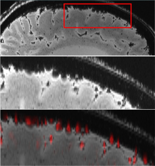
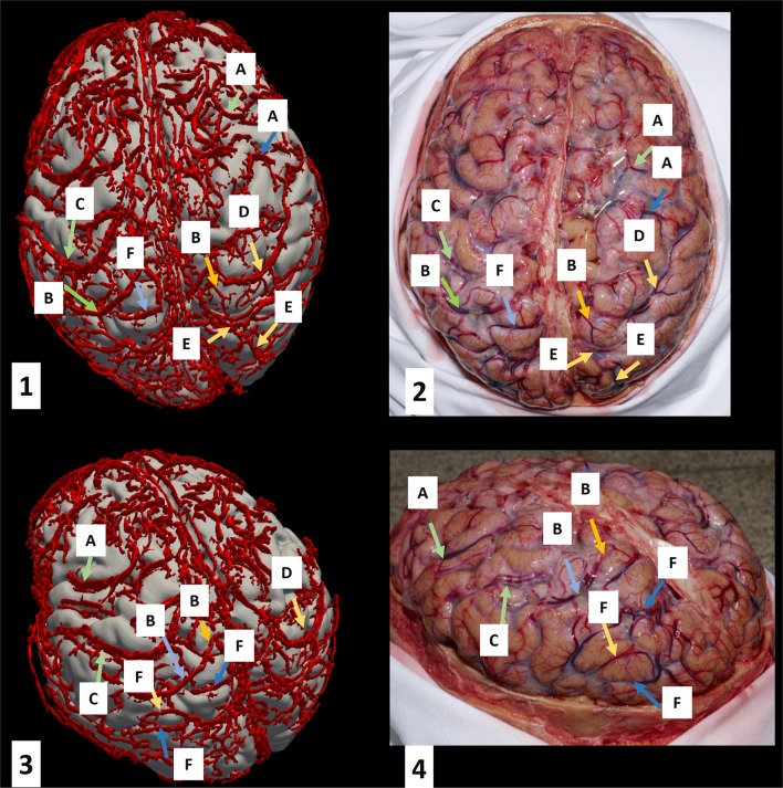
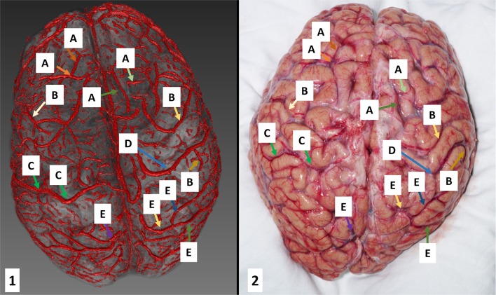
Image-guided neurosurgery uses information from a wide spectrum of methods to inform the neurosurgeon's judgement about which tissue to resect and which to spare. Imaging data are registered to the patient's skull so that they correspond to the intraoperative macro- and microscopic view. The correspondence between imaging and optical systems breaks down during surgery, however, as a result of cerebro-spinal fluid drain age, tissue resection, and gravity-based brain shift. In this work we investigate whether a map of surface veins, automatically segmented from MRI, could serve as additional reference system. Gradient-echo based [Formula: see text]-weighted imaging was performed on two human cadavers heads using a 7 Tesla MRI scanner. Automatic vessel segmentation was performed using the Frangi vesselness filter, and surface renderings of vessels compared with photographs of the surface of the brain following craniotomy. A high level of correspondence was established between vessel maps and the post autopsy photographs. Corresponding veins, including the prominent superior anastomotic veins, could be identified in all brain lobes. Automatic surface vessel segmentation is feasible and the high correspondence to post autopsy photographs indicates that they could be used as an additional reference system for image-guided neurosurgery in order to maintain the correspondence between imaging and optical systems.This has the advantage over a skull-based reference system that veins are clearly visible to the surgeon and move and deform with the underlying tissue, potentially making this surface net of landmarks robust to brain shift.
图像引导神经外科手术利用广泛的方法所提供的信息,来辅助神经外科医生判断切除哪些组织以及保留哪些组织。将成像数据与患者颅骨进行配准,以便使其与术中的宏观和微观视野相对应。然而,由于脑脊液引流、组织切除以及基于重力的脑移位,成像系统与光学系统之间的对应关系在手术过程中会被打破。在这项研究中,我们探究了从磁共振成像(MRI)中自动分割出来的脑表面静脉图谱是否可以作为一个额外的参考系统。使用一台7特斯拉的MRI扫描仪,对两具人类尸体头部进行了基于梯度回波的T2加权成像。利用Frangi血管造影滤波器进行血管自动分割,并将血管的表面渲染图与开颅术后的脑表面照片进行比较。在血管图谱与尸检照片之间建立了高度的对应关系。在所有脑叶中都能识别出相应的静脉,包括明显可见的上吻合静脉。自动脑表面血管分割是可行的,并且与尸检照片的高度对应表明,它们可以用作图像引导神经外科手术的额外参考系统,以维持成像系统与光学系统之间的对应关系。与基于颅骨的参考系统相比,这样做的优势在于,静脉对于外科医生来说清晰可见,并且会随着其下方的组织一起移动和变形,这可能使得这个表面标志网络对脑移位具有鲁棒性。