Ricci Alessandro, Di Vitantonio Hambra, De Paulis Danilo, Del Maestro Mattia, Raysi Soheila Dehcordi, Murrone Domenico, Luzzi Sabino, Galzio Renato Juan
Department of Neurosurgery, San Salvatore City Hospital, L'Aquila, Italy.
Department of Life, Health and Environmental Sciences (MESVA), University of L'Aquila, Italy.
Surg Neurol Int. 2017 Jun 13;8:117. doi: 10.4103/sni.sni_50_17. eCollection 2017.
Middle cerebral artery (MCA) aneurysms constitute from 18-40% of all intracranial aneurysms. They are mainly found in the proximal and bifurcation tracts and only in the 1.1-1.7% of cases they are located in the distal segment. The authors report a case of a ruptured saccular cortical MCA aneurysm with unknown etiology.
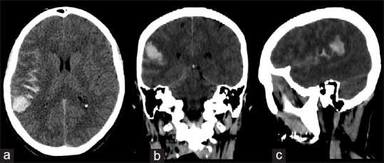
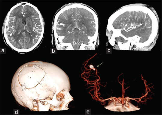
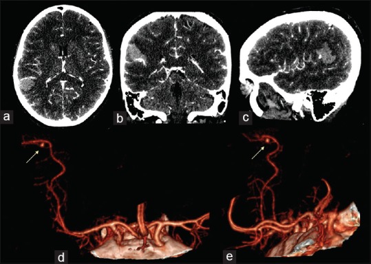
A 53-year-old female was admitted with a sudden severe headache, nausea, vomiting, and a slight left hemiparesis. The computed tomography (CT) scan showed subarachnoid hemorrhage (SAH) in the left sylvian fissure and intracerebral hemorrhage (ICH) in the left posterior parietal area. The CT angiography (CTA) reconstructed with 3D imaging showed a small saccular aneurysm in the M4 segment in proximity of the angular area. A left parieto-temporal craniotomy was performed, the aneurysm was clipped and the ICH evacuated. The motor deficit was progressively recovered. At 3-month follow-up examination, the patient was asymptomatic and feeling well.
In our opinion, surgery is the best choice for the treatment of ruptured M4 aneurysms with ICH, because it allows to evacuate the hematoma and to exclude the aneurysm from the intracranial circulation. In addition, we suggest both the use of the neuronavigation technique and of the indocyanine green videoangiography (ICGV) for the aneurismal surgery.
大脑中动脉(MCA)动脉瘤占所有颅内动脉瘤的18% - 40%。它们主要位于近端和分叉段,仅1.1% - 1.7%的病例位于远端段。作者报告一例病因不明的破裂囊状皮质MCA动脉瘤病例。
一名53岁女性因突发剧烈头痛、恶心、呕吐及轻度左侧偏瘫入院。计算机断层扫描(CT)显示左侧外侧裂蛛网膜下腔出血(SAH)及左侧顶叶后部脑出血(ICH)。经三维成像重建的CT血管造影(CTA)显示在角回区域附近的M4段有一个小囊状动脉瘤。行左侧颞顶开颅术,夹闭动脉瘤并清除脑出血。运动功能障碍逐渐恢复。在3个月的随访检查中,患者无症状且感觉良好。
我们认为,手术是治疗伴有脑出血的破裂M4动脉瘤的最佳选择,因为它既能清除血肿,又能将动脉瘤排除在颅内循环之外。此外,我们建议在动脉瘤手术中同时使用神经导航技术和吲哚菁绿视频血管造影(ICGV)。