Kesavan Gokul, Chekuru Avinash, Machate Anja, Brand Michael
Biotechnology Center and DFG-Research Center for Regenerative Therapies Dresden, Technische Universität DresdenDresden, Germany.
Front Neuroanat. 2017 Jun 30;11:52. doi: 10.3389/fnana.2017.00052. eCollection 2017.
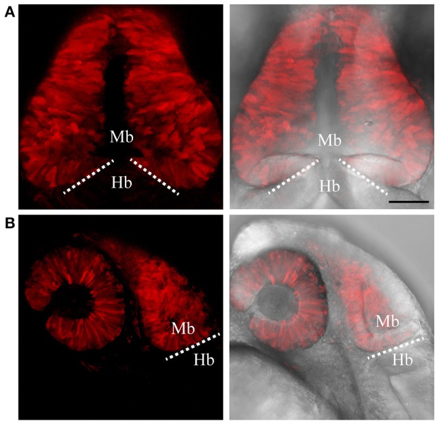
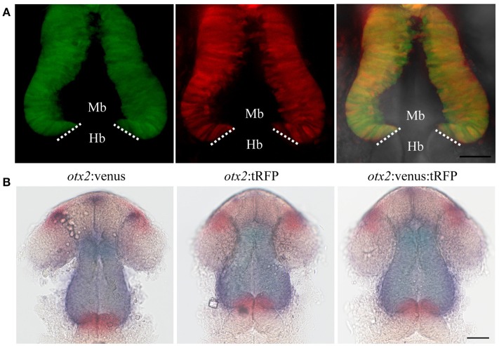
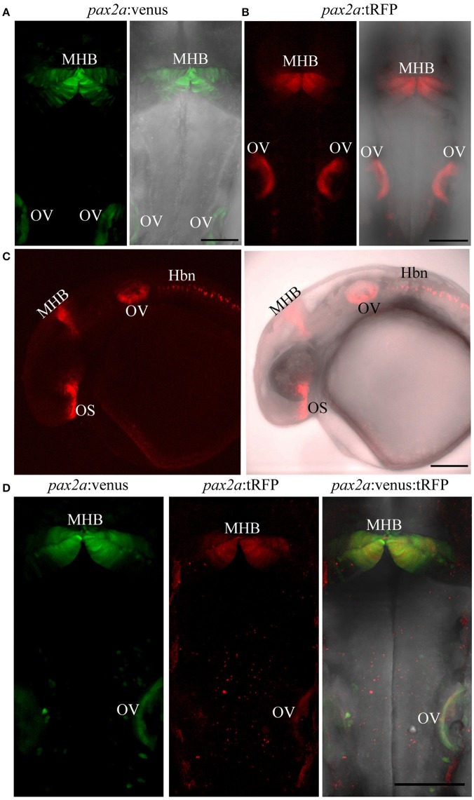
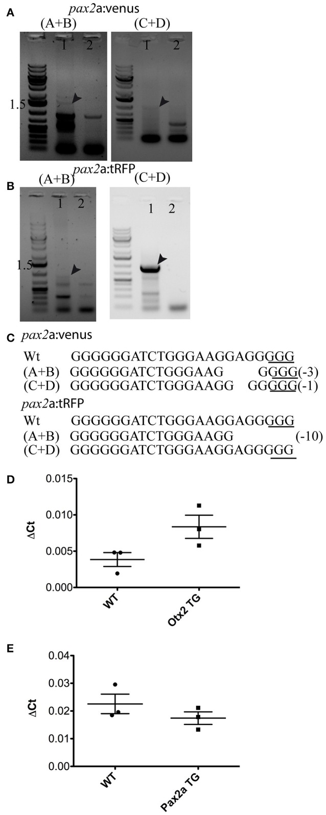
The midbrain-hindbrain boundary (MHB) acts as an organizer and controls the fate of neighboring cells to develop into either mesencephalic (midbrain) or metencephalic (hindbrain) cells by secreting signaling molecules like Wnt1 and Fgf8. The zebrafish is an excellent vertebrate model for studying MHB development due to the ease of gene manipulation and the possibility of following cellular dynamics and morphogenetic processes using live imaging. Currently, only very few reporter and/or Cre-driver lines are available to study gene expression at the MHB, hampering the understanding of MHB development, and traditional transgenic technologies using promoter/enhancer fragments or bacterial artificial chromosome (BAC)-mediated transgenesis often do not faithfully recapitulate endogenous expression patterns. In contrast, CRISPR/Cas9-mediated genome editing technology now provides a great opportunity to efficiently knock-in or knock-out genes. We have generated four CRISPR/Cas9-based knock-in fluorescent reporter lines for two crucial genes involved in MHB development, namely and . The coding sequences of the reporters were knocked-in upstream of the corresponding ATG and are, thus, under the control of the endogenous promoter/enhancer elements. Interestingly, this strategy does not disturb endogenous gene expression. Using the fast maturing fluorescent protein reporter, Venus, enabled us to follow MHB development using cell tracking and live imaging. In addition, we show that these reporter lines label various neuronal and glial cell types in the adult zebrafish brain, making them highly suitable for investigating embryonic and adult midbrain, hindbrain, and MHB development.
中脑-后脑边界(MHB)作为一个组织者,通过分泌如Wnt1和Fgf8等信号分子,控制邻近细胞的命运,使其发育成中脑或后脑细胞。斑马鱼是研究MHB发育的优秀脊椎动物模型,因为其基因操作简便,且可以利用活体成像追踪细胞动力学和形态发生过程。目前,仅有极少数报告基因和/或Cre驱动系可用于研究MHB处的基因表达,这阻碍了对MHB发育的理解,并且使用启动子/增强子片段或细菌人工染色体(BAC)介导的转基因的传统转基因技术往往不能忠实地重现内源性表达模式。相比之下,CRISPR/Cas9介导的基因组编辑技术现在为高效敲入或敲除基因提供了一个绝佳机会。我们已经为两个参与MHB发育的关键基因生成了四个基于CRISPR/Cas9的敲入荧光报告系,即 和 。报告基因的编码序列被敲入相应ATG的上游,因此受内源性启动子/增强子元件的控制。有趣的是,这种策略不会干扰内源性基因表达。使用快速成熟的荧光蛋白报告基因Venus,使我们能够通过细胞追踪和活体成像来追踪MHB的发育。此外,我们表明这些报告系标记了成年斑马鱼脑中的各种神经元和神经胶质细胞类型,使其非常适合研究胚胎期和成年期的中脑、后脑以及MHB的发育。