Gibbs Holly C, Chang-Gonzalez Ana, Hwang Wonmuk, Yeh Alvin T, Lekven Arne C
Department of Biomedical Engineering, Texas A&M UniversityCollege Station, TX, United States.
Department of Materials Science and Engineering, Texas A&M UniversityCollege Station, TX, United States.
Front Neuroanat. 2017 Aug 3;11:64. doi: 10.3389/fnana.2017.00064. eCollection 2017.
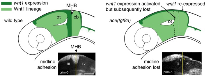
A constriction in the neural tube at the junction of the midbrain and hindbrain is a conserved feature of vertebrate embryos. The constriction is a defining feature of the midbrain-hindbrain boundary (MHB), a signaling center that patterns the adjacent midbrain and rostral hindbrain and forms at the junction of two gene expression domains in the early neural plate: an anterior positive domain and a posterior positive domain. and genes encode mutually repressive transcription factors that create a lineage restriction boundary at their expression interface. Wnt and Fgf genes form a mutually dependent feedback system that maintains their expression domains on the or side of the boundary, respectively. Constriction morphogenesis occurs after these conserved gene expression domains are established and while their mutual interactions maintain their expression pattern; consequently, mutant studies in zebrafish have led to the suggestion that constriction morphogenesis should be considered a unique phase of MHB development. We analyzed MHB morphogenesis in loss of function zebrafish embryos using a reporter driven by the conserved enhancer to visualize anterior boundary cells. We found that loss of function results in a re-activation of reporter expression posterior to the boundary simultaneous with an inactivation of the reporter in the anterior boundary cells, and that these events correlate with relaxation of the boundary constriction. In consideration of other results that correlate the boundary constriction with Wnt and Fgf expression, we propose that the maintenance of an active Wnt-Fgf feedback loop is a key factor in driving the morphogenesis of the MHB constriction.
中脑与后脑交界处神经管的收缩是脊椎动物胚胎的一个保守特征。这种收缩是中脑-后脑边界(MHB)的一个决定性特征,MHB是一个信号中心,它决定相邻中脑和后脑前部的模式,并在早期神经板的两个基因表达域的交界处形成:一个前部阳性域和一个后部阳性域。 和 基因编码相互抑制的转录因子,它们在其表达界面处形成一个谱系限制边界。Wnt和Fgf基因形成一个相互依赖的反馈系统,分别在边界的 或 侧维持它们的表达域。收缩形态发生在这些保守的基因表达域建立之后,并且在它们的相互作用维持其表达模式时发生;因此,斑马鱼的突变研究表明,收缩形态发生应被视为MHB发育的一个独特阶段。我们使用由保守的 增强子驱动的报告基因来可视化前部边界细胞,分析了功能缺失的斑马鱼胚胎中的MHB形态发生。我们发现,功能缺失导致边界后方的 报告基因表达重新激活,同时前部边界细胞中的 报告基因失活,并且这些事件与边界收缩的松弛相关。考虑到其他将边界收缩与Wnt和Fgf表达相关联的结果,我们提出维持活跃的Wnt-Fgf反馈环是驱动MHB收缩形态发生的关键因素。