Department of Internal Medicine, Guizhou Osteological Hospital, Guiyang, Guizhou 550007, P.R. China.
Department of Pathology, Guizhou Osteological Hospital, Guiyang, Guizhou 550007, P.R. China.
Mol Med Rep. 2017 Oct;16(4):3958-3964. doi: 10.3892/mmr.2017.7064. Epub 2017 Jul 21.
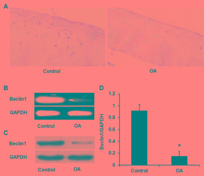
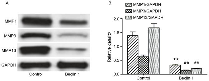
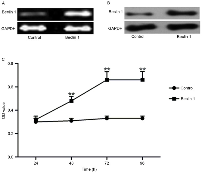
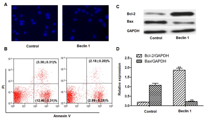
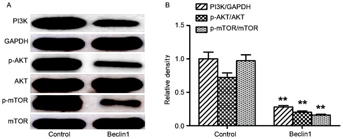
In the present study, the expression of Beclin 1 in osteoarthritis (OA) cartilage tissue was investigated, and also its role in proliferation, apoptosis and expression of matrix metalloproteinases (MMPs) in chondrocytes obtained from patients with OA. Beclin 1 expression in cartilage tissue from OA patients, and in the age- and sex-matched controls, was detected by immunohistochemistry, semi-quantitative polymerase chain reaction and western blotting. Chondrocytes were divided into control and Beclin 1-overexpressed groups. After transfection for 48, 72 and 96 h, cell viability, apoptosis, the phosphatidylinositol-3-kinase (PI3K)/protein kinase B (Akt)/mammalian target of rapamycin (mTOR) signaling pathway and MMPs were examined. The mRNA and protein expression levels of Beclin 1 were significantly decreased in cartilage tissue from OA patients compared with the sex- and age-matched controls (P<0.05). In chondrocytes from OA patients, Beclin 1 overexpression significantly increased cell viability (P<0.05). Beclin 1 overexpression additionally decreased the degree of apoptosis, as demonstrated by Hoechst staining and flow cytometric analysis. B-cell lymphoma-2 (Bcl-2) was upregulated, and Bcl-2 associated X was downregulated, following Beclin 1 overexpression (P<0.05). The PI3K/Akt/mTOR signaling pathway was mitigated following Beclin 1 overexpression (P<0.05). In addition, MMP1, MMP3 and MMP13 were downregulated after Beclin 1 overexpression (P<0.05). Taken together, low expression levels of Beclin 1 may contribute towards the degeneration of chondrocytes. Beclin 1 overexpression increased cell viability, inhibited apoptosis and MMPs, likely via the PI3K/Akt/mTOR signaling pathway.
在本研究中,研究了自噬相关蛋白 Beclin 1 在骨关节炎(OA)软骨组织中的表达及其在 OA 患者软骨细胞增殖、凋亡和基质金属蛋白酶(MMPs)表达中的作用。采用免疫组织化学、半定量聚合酶链反应和 Western blot 检测 OA 患者和年龄、性别匹配的对照组软骨组织中 Beclin 1 的表达。将软骨细胞分为对照组和 Beclin 1 过表达组。转染 48、72 和 96 h 后,检测细胞活力、凋亡、磷酸肌醇 3-激酶(PI3K)/蛋白激酶 B(Akt)/哺乳动物雷帕霉素靶蛋白(mTOR)信号通路和 MMPs。与性别和年龄匹配的对照组相比,OA 患者软骨组织中 Beclin 1 的 mRNA 和蛋白表达水平明显降低(P<0.05)。在 OA 患者的软骨细胞中,Beclin 1 过表达显著增加了细胞活力(P<0.05)。Beclin 1 过表达还降低了 Hoechst 染色和流式细胞术分析显示的凋亡程度。Beclin 1 过表达后,B 细胞淋巴瘤-2(Bcl-2)上调,Bcl-2 相关 X 下调(P<0.05)。Beclin 1 过表达后,PI3K/Akt/mTOR 信号通路被抑制(P<0.05)。此外,Beclin 1 过表达后 MMP1、MMP3 和 MMP13 下调(P<0.05)。总之,Beclin 1 低表达可能导致软骨细胞退变。Beclin 1 过表达通过 PI3K/Akt/mTOR 信号通路增加细胞活力,抑制凋亡和 MMPs。