Amparo Francisco, Yin Jia, Di Zazzo Antonio, Abud Tulio, Jurkunas Ula V, Hamrah Pedram, Dana Reza
Massachusetts Eye and Ear Infirmary, Department of Ophthalmology, Harvard Medical School, Boston, MA, USA.
Cornea Service, New England Eye Center, Department of Ophthalmology, Tufts Medical Center, Tufts University School of Medicine, Boston, MA, USA.
Transl Vis Sci Technol. 2017 Jul 20;6(4):13. doi: 10.1167/tvst.6.4.13. eCollection 2017 Jul.
To evaluate interobserver concordance in measured ocular redness among a group of raters using an objective computer-assisted method (ocular redness index [ORI]) and a group of clinicians using an ordinal comparative scale.
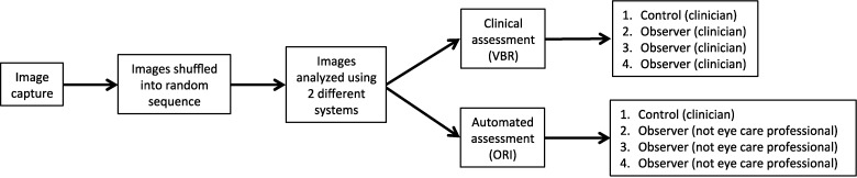
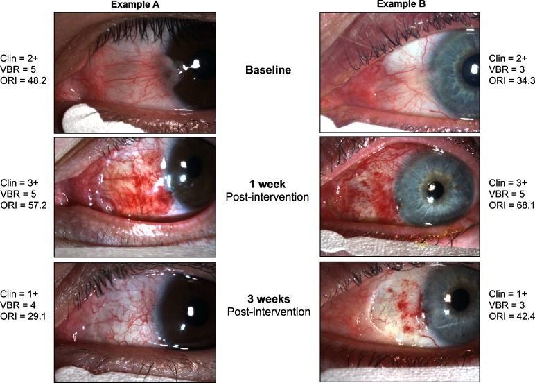
We conducted a prospective study to evaluate ocular redness in clinical photographs of 12 patients undergoing pterygium surgery. Photographs were acquired preoperatively, and at 1 week and 1 month postoperatively. One group of clinicians graded conjunctival redness in the photographs using an image-based comparative scale. A second group applied the ORI to measure redness in the same photographs. We evaluated redness change between time points, level of agreement among raters, and assessed redness score differences among observers within each group.
Interobserver agreement using the image-based redness scale was 0.458 ( < 0.001). Interobserver agreement with the ORI was 0.997 ( < 0.001). We observed statistically significant differences among clinicians' measurements obtained with the image-based redness scale ( < 0.001). There were no significant differences among measurements obtained with the ORI ( = 0.27). We observed a significant change in redness between baseline and follow-up visits with all scoring methods. Detailed analysis of redness change was performed only in the ORI group due to availability of continuous scores.
Our findings suggest that the ORI scores provide higher consistency among raters than ordinal scales, and can discriminate redness changes that clinical observers often can miss.
The ORI may be a reliable alternative to measure ocular redness objectively in the clinic and in clinical trials.
使用客观的计算机辅助方法(眼部发红指数[ORI])评估一组评估者之间在测量眼部发红方面的观察者间一致性,并使用序数比较量表评估一组临床医生之间的一致性。
我们进行了一项前瞻性研究,以评估12例接受翼状胬肉手术患者临床照片中的眼部发红情况。在术前、术后1周和术后1个月采集照片。一组临床医生使用基于图像的比较量表对照片中的结膜发红进行分级。另一组使用ORI测量相同照片中的发红情况。我们评估了不同时间点之间的发红变化、评估者之间的一致性水平,并评估了每组内观察者之间的发红评分差异。
使用基于图像的发红量表的观察者间一致性为0.458(<0.001)。使用ORI的观察者间一致性为0.997(<0.001)。我们观察到临床医生使用基于图像的发红量表获得的测量结果之间存在统计学显著差异(<0.001)。使用ORI获得的测量结果之间没有显著差异(=0.27)。我们观察到所有评分方法在基线和随访之间的发红有显著变化。由于可获得连续评分,仅在ORI组中对发红变化进行了详细分析。
我们的研究结果表明,与序数量表相比,ORI评分在评估者之间提供了更高的一致性,并且可以区分临床观察者经常错过的发红变化。
ORI可能是在临床和临床试验中客观测量眼部发红的可靠替代方法。