Brezavšček Miha, Fawzy Ahmed, Bächle Maria, Tuna Taskin, Fischer Jens, Att Wael
Department of Prosthodontics, School of Dentistry, Albert-Ludwigs University, Hugstetter Strasse 55, 79106 Freiburg, Germany.
Institute for Dental Materials and Engineering, University Hospital for Dental Medicine, University of Basel, 4056 Basel, Switzerland.
Materials (Basel). 2016 Nov 24;9(12):958. doi: 10.3390/ma9120958.
Improvements in the bioactivity of zirconia implants for accelerated healing and reduced morbidity have been of continuing interest in the fields of dentistry and orthopedic surgery. The aim of the present study was to examine whether UV treatment increases the osteoconductivity of zirconia-based materials.
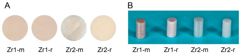
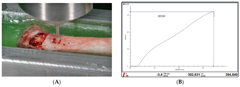
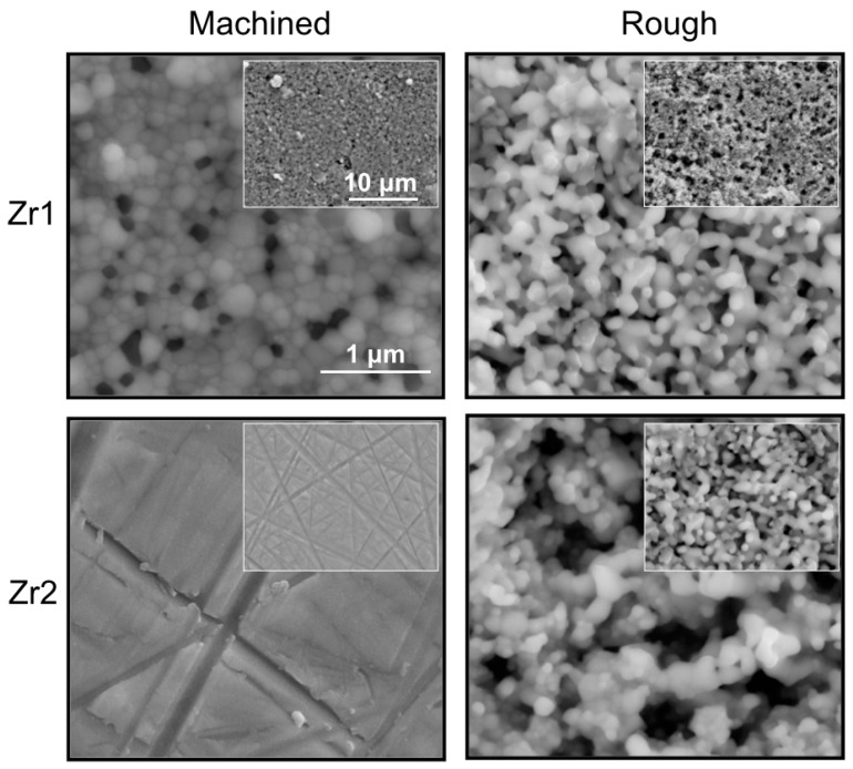
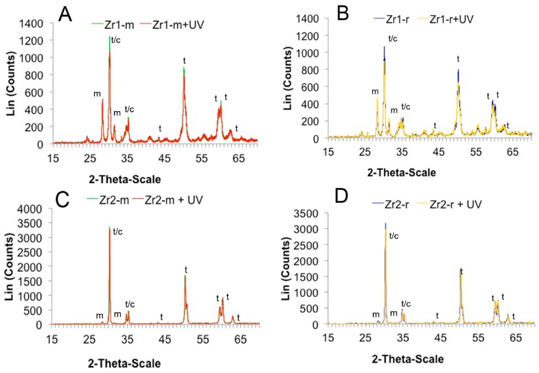
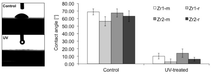
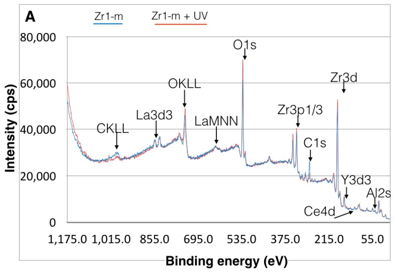
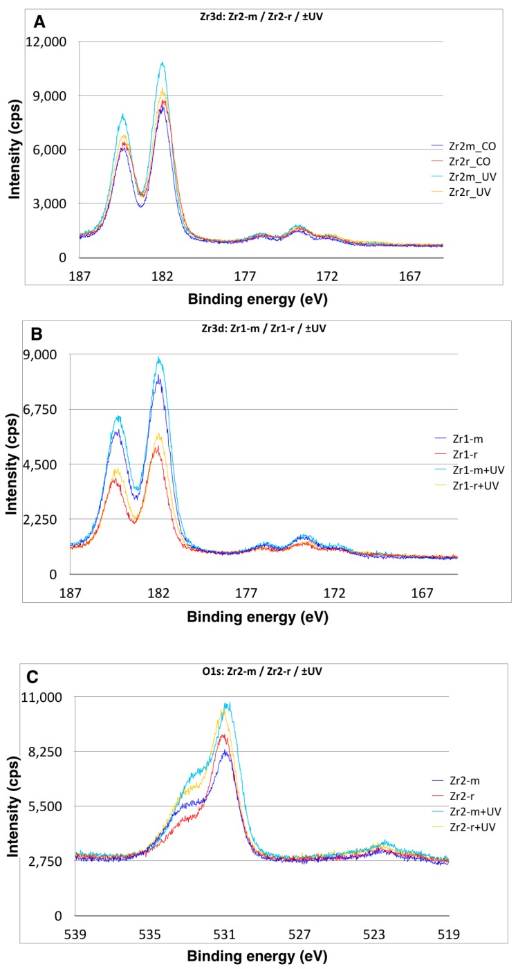
Smooth and rough zirconia-based disks and cylindrical implants were treated with UV light for 15 min and subsequently placed in rat femurs. Surface characterization was performed using scanning electron microscopy (SEM), atomic force microscopy (AFM), X-ray photoelectron spectroscopy (XPS) and contact angle measurements.
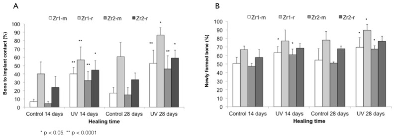
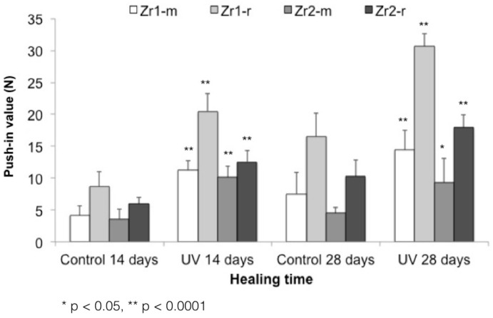
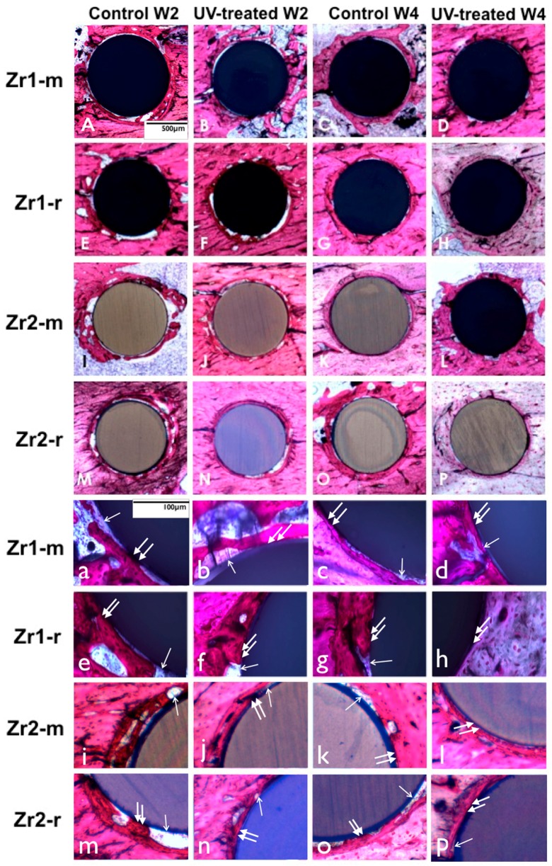
In vivo histomorphometry revealed that the percentage of bone-implant contact and the amount of bone volume, formed around UV-treated implants, increased by 3-7-fold for smooth surfaces and by 1.4-1.7-fold for rough surfaces compared to non-treated specimens at Weeks 2 and 4 of healing, respectively. A biomechanical test showed that UV treatment accelerated the establishment of bone-zirconia integration and enhanced the strength of the bone-implant interface by two-fold. Additionally, surface characterization of the zirconia disks revealed that UV treatment decreased the amount of surface carbon and converted the hydrophilic status from hydrophobic to superhydrophilic.
This study indicates that UV light pretreatment enhances the osteoconductive capacity of zirconia-based materials.
提高氧化锆植入物的生物活性以加速愈合并降低发病率,一直是牙科和整形外科领域持续关注的问题。本研究的目的是检验紫外线处理是否能提高氧化锆基材料的骨传导性。
将光滑和粗糙的氧化锆基圆盘及圆柱形植入物用紫外线处理15分钟,随后植入大鼠股骨。使用扫描电子显微镜(SEM)、原子力显微镜(AFM)、X射线光电子能谱(XPS)和接触角测量进行表面表征。
体内组织形态计量学显示,在愈合的第2周和第4周,与未处理的标本相比,紫外线处理过的植入物周围形成的骨-植入物接触百分比和骨体积量,光滑表面增加了3至7倍,粗糙表面增加了1.4至1.7倍。生物力学测试表明,紫外线处理加速了骨与氧化锆的结合,并使骨-植入物界面的强度提高了两倍。此外,氧化锆圆盘的表面表征显示,紫外线处理减少了表面碳含量,并将亲水性状态从疏水性转变为超亲水性。
本研究表明,紫外线预处理可增强氧化锆基材料的骨传导能力。