Yonsei University, School of Mechanical Engineering, Seoul, 03722, Republic of Korea.
Yonsei University, School of Integrated Technology & Yonsei Institute of Convergence Technology, Incheon, 21983, Republic of Korea.
Sci Rep. 2017 Aug 8;7(1):7564. doi: 10.1038/s41598-017-07703-w.
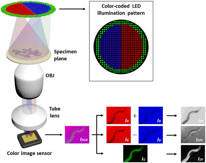
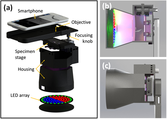
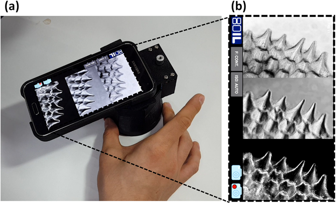
We present a portable multi-contrast microscope capable of producing bright-field, dark-field, and differential phase contrast images of thin biological specimens on a smartphone platform. The microscopy method is based on an imaging scheme termed "color-coded light-emitting-diode (LED) microscopy (cLEDscope)," in which a specimen is illuminated with a color-coded LED array and light transmitted through the specimen is recorded by a color image sensor. Decomposition of the image into red, green, and blue colors and subsequent computation enable multi-contrast imaging in a single shot. In order to transform a smartphone into a multi-contrast imaging device, we developed an add-on module composed of a patterned color micro-LED array, specimen stage, and miniature objective. Simple installation of this module onto a smartphone enables multi-contrast imaging of transparent specimens. In addition, an Android-based app was implemented to acquire an image, perform the associated computation, and display the multi-contrast images in real time. Herein, the details of our smartphone module and experimental demonstrations with various biological specimens are presented.
我们展示了一种便携式多对比度显微镜,能够在智能手机平台上对薄生物样本产生明场、暗场和相差对比图像。该显微镜方法基于一种称为“彩色发光二极管 (LED) 显微镜 (cLEDscope) 的成像方案”,其中用彩色 LED 阵列照明样本,通过彩色图像传感器记录通过样本的光。将图像分解为红色、绿色和蓝色,然后进行计算,可在单次拍摄中实现多对比度成像。为了将智能手机转变为多对比度成像设备,我们开发了一个附加模块,由图案化彩色微 LED 阵列、样本台和微型物镜组成。将该模块简单地安装到智能手机上,即可对透明样本进行多对比度成像。此外,还实现了一个基于 Android 的应用程序,用于获取图像、执行相关计算,并实时显示多对比度图像。本文介绍了我们的智能手机模块的详细信息以及各种生物样本的实验演示。