Deryagin Oleg G, Gavrilova Svetlana A, Gainutdinov Khalil L, Golubeva Anna V, Andrianov Vyatcheslav V, Yafarova Guzel G, Buravkov Sergey V, Koshelev Vladimir B
Department of Physiology and General Pathology, Medical Faculty, Lomonosov Moscow State UniversityMoscow, Russia.
Laboratory of Neurorehabilitation of Motor Disorders, Institute of Fundamental Medicine and Biology, Kazan Federal UniversityKazan, Russia.
Front Neurosci. 2017 Jul 25;11:427. doi: 10.3389/fnins.2017.00427. eCollection 2017.
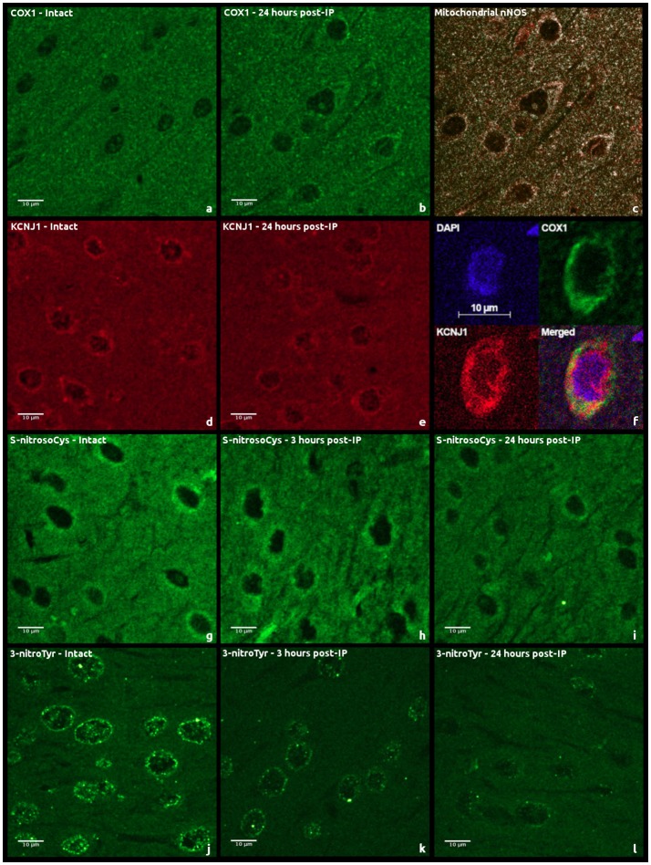
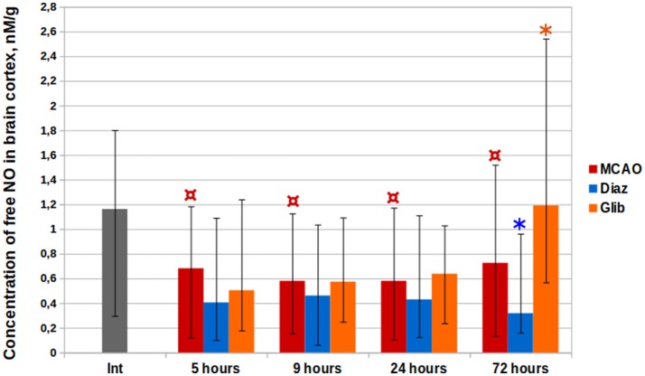
Preconditioning of the brain induces tolerance to the damaging effects of ischemia and prevents cell death in ischemic penumbra. The development of this phenomenon is mediated by mitochondrial adenosine triphosphate-sensitive potassium ([Formula: see text]) channels and nitric oxide signaling (NO). The aim of this study was to investigate the dynamics of molecular changes in mitochondria after ischemic preconditioning (IP) and the effect of pharmacological preconditioning (PhP) with the [Formula: see text]-channels opener diazoxide on NO levels after ischemic stroke in rats. Immunofluorescence-histochemistry and laser-confocal microscopy were applied to evaluate the cortical expression of electron transport chain enzymes, mitochondrial [Formula: see text]-channels, neuronal and inducible NO-synthases, as well as the dynamics of nitrosylation and nitration of proteins in rats during the early and delayed phases of IP. NO cerebral content was studied with electron paramagnetic resonance (EPR) spectroscopy using spin trapping. We found that 24 h after IP in rats, there is a two-fold decrease in expression of mitochondrial [Formula: see text]-channels ( = 0.012) in nervous tissue, a comparable increase in expression of cytochrome c oxidase ( = 0.008), and a decrease in intensity of protein S-nitrosylation and nitration ( = 0.0004 and = 0.001, respectively). PhP led to a 56% reduction of free NO concentration 72 h after ischemic stroke simulation ( = 0.002). We attribute this result to the restructuring of tissue energy metabolism, namely the provision of increased catalytic sites to mitochondria and the increased elimination of NO, which prevents a decrease in cell sensitivity to oxygen during subsequent periods of severe ischemia.
脑预处理可诱导对缺血性损伤的耐受性,并防止缺血半暗带中的细胞死亡。这一现象的发生是由线粒体三磷酸腺苷敏感性钾([公式:见原文])通道和一氧化氮信号传导(NO)介导的。本研究的目的是探讨缺血预处理(IP)后线粒体分子变化的动态过程,以及用[公式:见原文]通道开放剂二氮嗪进行药物预处理(PhP)对大鼠缺血性卒中后NO水平的影响。应用免疫荧光组织化学和激光共聚焦显微镜评估电子传递链酶、线粒体[公式:见原文]通道、神经元型和诱导型NO合酶在皮质的表达,以及IP早期和延迟期大鼠蛋白质亚硝化和硝化的动态变化。使用自旋捕获技术通过电子顺磁共振(EPR)光谱研究脑内NO含量。我们发现,大鼠IP后24小时,神经组织中线粒体[公式:见原文]通道的表达下降了两倍(=0.012),细胞色素c氧化酶的表达有类似程度的增加(=0.008),蛋白质S-亚硝化和硝化的强度降低(分别为=0.0004和=0.001)。PhP导致缺血性卒中模拟后72小时游离NO浓度降低56%(=0.002)。我们将这一结果归因于组织能量代谢的重构,即线粒体催化位点增加以及NO清除增加,这可防止在随后的严重缺血期细胞对氧的敏感性降低。