Mighri Nabila, Mao Jifu, Mighri Frej, Ajji Abdallah, Rouabhia Mahmoud
Groupe de Recherche en Écologie Buccale, Faculté de Médecine Dentaire, Université Laval, 2420 rue de la Terrasse, Québec, QC G1V 0A6, Canada.
Department of Chemical Engineering, Université Laval, 1065 avenue de la Médecine, Québec, QC G1V 0A6, Canada.
Materials (Basel). 2015 Nov 13;8(11):7673-7689. doi: 10.3390/ma8115413.
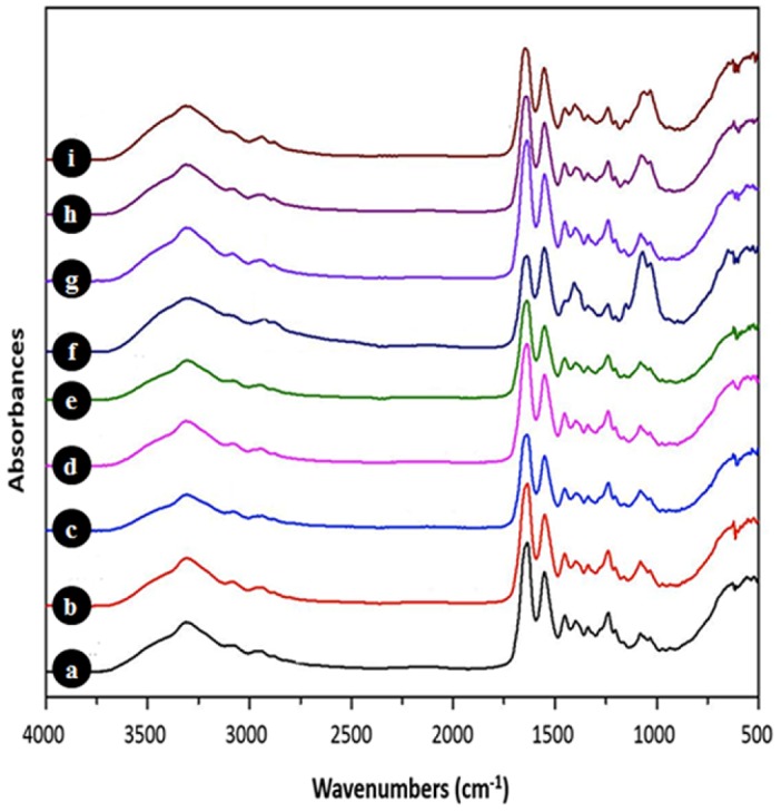
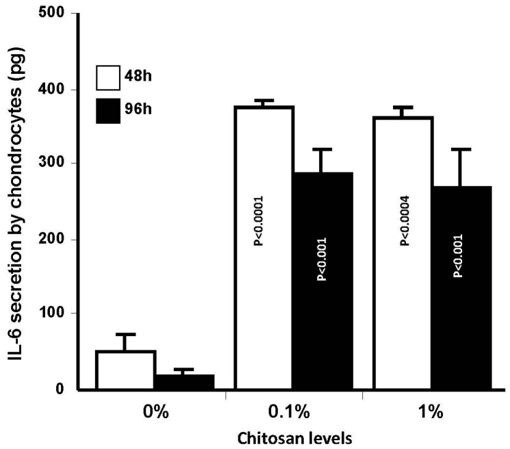
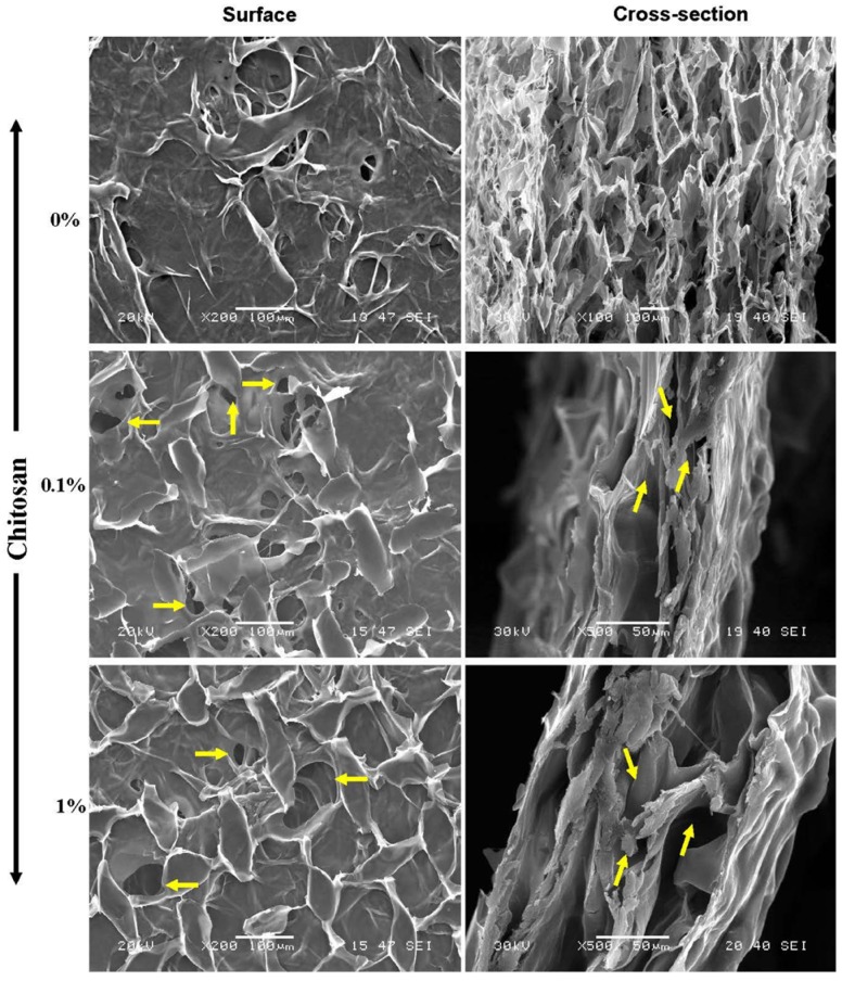
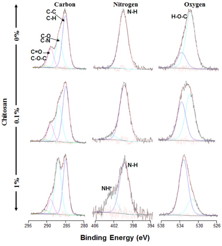
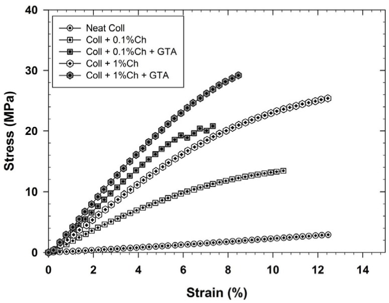
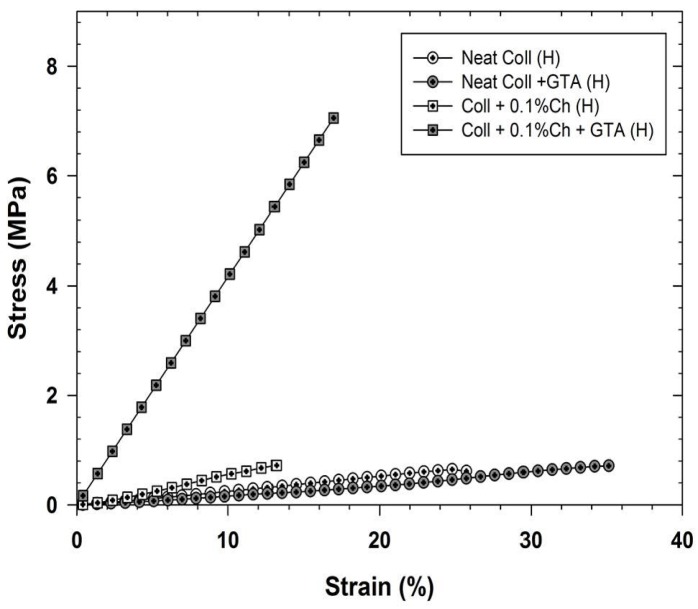
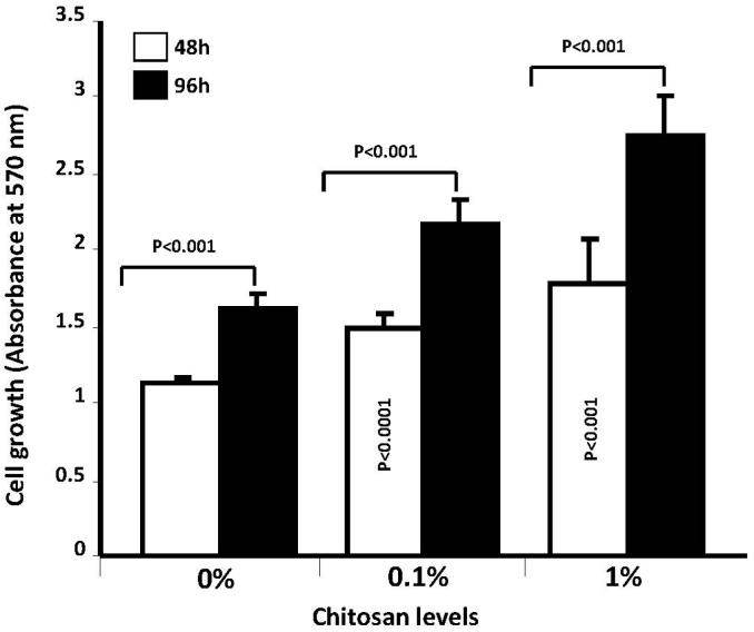
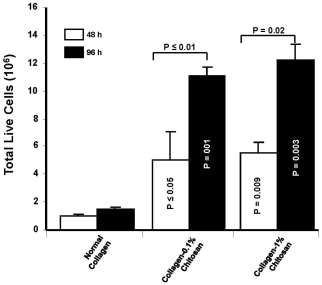
Designing scaffolds made from natural polymers may be highly attractive for tissue engineering strategies. We sought to produce and characterize chitosan-coated collagen membranes and to assess their efficacy in promoting chondrocyte adhesion, growth, and cytokine secretion. Porous collagen membranes were placed in chitosan solutions then crosslinked with glutaraldehyde vapor. Fourier transform infrared (FTIR) analyses showed elevated absorption at 1655 cm of the carbon-nitrogen (N=C) bonds formed by the reaction between the (NH₂) of the chitosan and the (C=O) of the glutaraldehyde. A significant peak in the amide II region revealed a significant deacetylation of the chitosan. Scanning electron microscopy (SEM) images of the chitosan-coated membranes exhibited surface variations, with pore size ranging from 20 to 50 µm. X-ray photoelectron spectroscopy (XPS) revealed a decreased C-C groups and an increased C-N/C-O groups due to the reaction between the carbon from the collagen and the NH2 from the chitosan. Increased rigidity of these membranes was also observed when comparing the chitosan-coated and uncoated membranes at dried conditions. However, under wet conditions, the chitosan coated collagen membranes showed lower rigidity as compared to dried conditions. Of great interest, the glutaraldehyde-crosslinked chitosan-coated collagen membranes promoted chondrocyte adhesion, growth, and interleukin (IL)-6 secretion. Overall results confirm the feasibility of using designed chitosan-coated collagen membranes in future applications, such as cartilage repair.
设计由天然聚合物制成的支架对于组织工程策略可能极具吸引力。我们试图制备并表征壳聚糖包被的胶原膜,并评估其在促进软骨细胞黏附、生长和细胞因子分泌方面的功效。将多孔胶原膜置于壳聚糖溶液中,然后用戊二醛蒸汽交联。傅里叶变换红外光谱(FTIR)分析显示,壳聚糖的(NH₂)与戊二醛的(C=O)反应形成的碳氮(N=C)键在1655 cm处的吸收增加。酰胺II区域的一个显著峰表明壳聚糖发生了显著的脱乙酰化。壳聚糖包被膜的扫描电子显微镜(SEM)图像显示出表面变化,孔径范围为20至50 µm。X射线光电子能谱(XPS)显示,由于胶原中的碳与壳聚糖中的NH2反应,C-C基团减少,C-N/C-O基团增加。在干燥条件下比较壳聚糖包被膜和未包被膜时,还观察到这些膜的刚性增加。然而,在潮湿条件下,壳聚糖包被的胶原膜与干燥条件相比显示出较低的刚性。非常有趣的是,戊二醛交联的壳聚糖包被胶原膜促进了软骨细胞的黏附、生长和白细胞介素(IL)-6的分泌。总体结果证实了在未来应用中,如软骨修复,使用设计的壳聚糖包被胶原膜的可行性。