Yasuda Keisuke, Okazaki Yohei, Abe Yasuhiko, Tsuga Kazuhiro
Department of Advanced Prosthodontics, Division of Dental Sciences, Biomedical Sciences Major, Graduate School of Biomedical & Health Sciences, Hiroshima University, 1-2-3 Kasumi, Minami-ku, Hiroshima 734-8553, Japan.
Heliyon. 2017 Aug 1;3(8):e00372. doi: 10.1016/j.heliyon.2017.e00372. eCollection 2017 Aug.
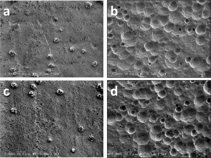
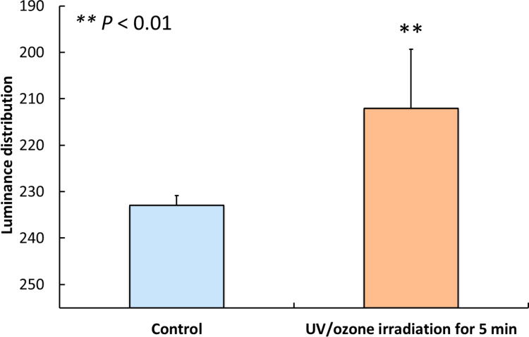
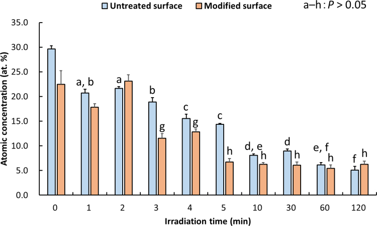
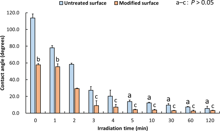
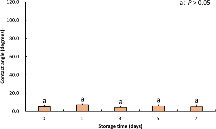
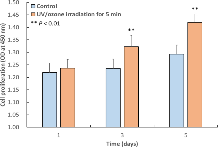
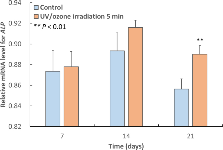
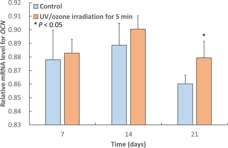
The purpose of this study was to establish whether UV/ozone (O) irradiation method can effectively decontaminate hydroxyapatite surfaces, including those modified by the treatment with 30% phosphoric acid solution through morphological and chemical surface analyses (surface roughness, X-ray photoelectron spectroscopy and wettability), and to evaluate the response of osteoblast-like MC3T3-E1 cells to the modified hydroxyapatite surface decontaminated via this method. The amount of carbon and the contact angle of hydroxyapatite surfaces were significantly decreased by UV/O irradiation that lasted for ≥ 5 and ≥ 3 min, respectively ( < 0.01). Additionally, 7-day storage of HPO-modified hydroxyapatite surface decontaminated with 5-min irradiation did not affect contact angle values ( > 0.05). MC3T3-E1 cell proliferation, differentiation (as assessed by relative and mRNA levels), and mineralisation were significantly promoted on irradiated surfaces ( < 0.05). These findings show that UV/O irradiation for ≥ 5 min significantly decontaminated HPO-modified hydroxyapatite surface, improved its wettability, and facilitated osteoblast growth and function.
本研究的目的是通过形态学和化学表面分析(表面粗糙度、X射线光电子能谱和润湿性)确定紫外线/臭氧(UV/O)辐照方法是否能有效净化羟基磷灰石表面,包括经30%磷酸溶液处理改性的表面,并评估成骨样MC3T3-E1细胞对通过该方法净化的改性羟基磷灰石表面的反应。UV/O辐照分别持续≥5分钟和≥3分钟后,羟基磷灰石表面的碳含量和接触角显著降低(P<0.01)。此外,经5分钟辐照净化的HPO改性羟基磷灰石表面储存7天不影响接触角值(P>0.05)。在辐照表面上,MC3T3-E1细胞的增殖、分化(通过相对ALP和Runx mRNA水平评估)和矿化显著促进(P<0.05)。这些结果表明,UV/O辐照≥5分钟可显著净化HPO改性羟基磷灰石表面,改善其润湿性,并促进成骨细胞生长和功能。