• 文献检索
  • 文档翻译
  • 深度研究
  • 学术资讯
  • Suppr Zotero 插件Zotero 插件
  • 邀请有礼
  • 套餐&价格
  • 历史记录
应用&插件
Suppr Zotero 插件Zotero 插件浏览器插件Mac 客户端Windows 客户端微信小程序
定价
高级版会员购买积分包购买API积分包
服务
文献检索文档翻译深度研究API 文档MCP 服务
关于我们
关于 Suppr公司介绍联系我们用户协议隐私条款
关注我们

Suppr 超能文献

核心技术专利:CN118964589B侵权必究
粤ICP备2023148730 号-1Suppr @ 2026

文献检索

告别复杂PubMed语法,用中文像聊天一样搜索,搜遍4000万医学文献。AI智能推荐,让科研检索更轻松。

立即免费搜索

文件翻译

保留排版,准确专业,支持PDF/Word/PPT等文件格式,支持 12+语言互译。

免费翻译文档

深度研究

AI帮你快速写综述,25分钟生成高质量综述,智能提取关键信息,辅助科研写作。

立即免费体验

通过高内涵成像评估生物相容性氧化铁纳米颗粒与细胞的相互作用,这些纳米颗粒可用作磁共振成像的潜在对比剂。

Assessing cell-nanoparticle interactions by high content imaging of biocompatible iron oxide nanoparticles as potential contrast agents for magnetic resonance imaging.

机构信息

Biophysics Group, Department of Physics and Astronomy, University College London, Gower Street, London, WC1E 6BT, UK.

UCL Healthcare and Biomagnetics and Nanomaterials Laboratory, 21 Albemarle Street, London, W1S 4BS, UK.

出版信息

Sci Rep. 2017 Aug 10;7(1):7850. doi: 10.1038/s41598-017-08092-w.

DOI:10.1038/s41598-017-08092-w
PMID:28798327
原文链接:https://pmc.ncbi.nlm.nih.gov/articles/PMC5552868/
Abstract

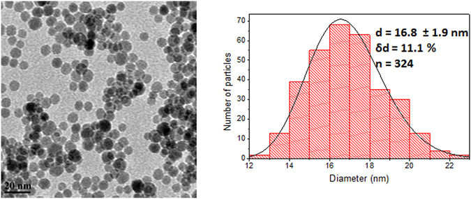
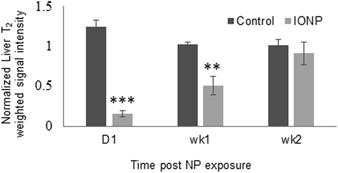
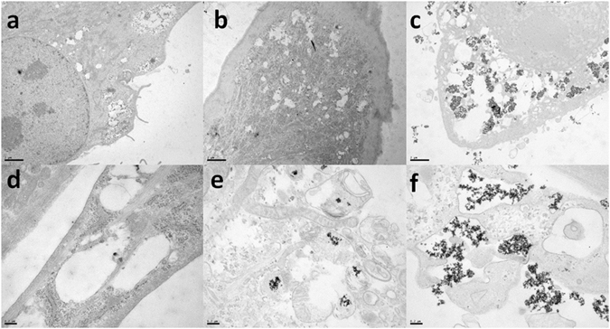
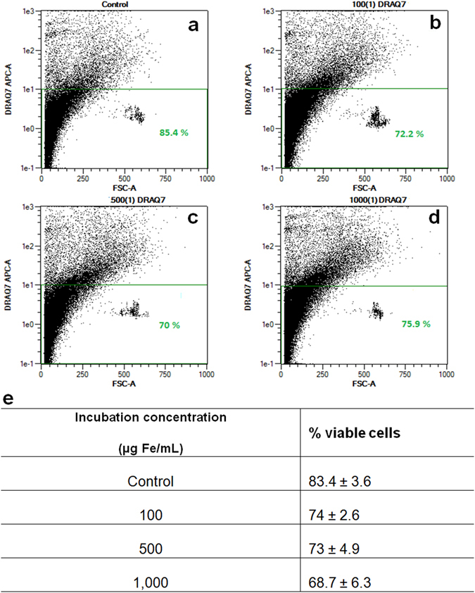
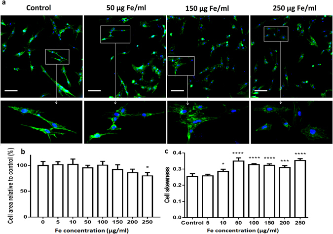
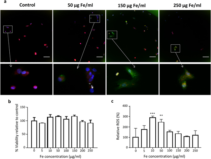
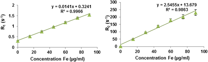
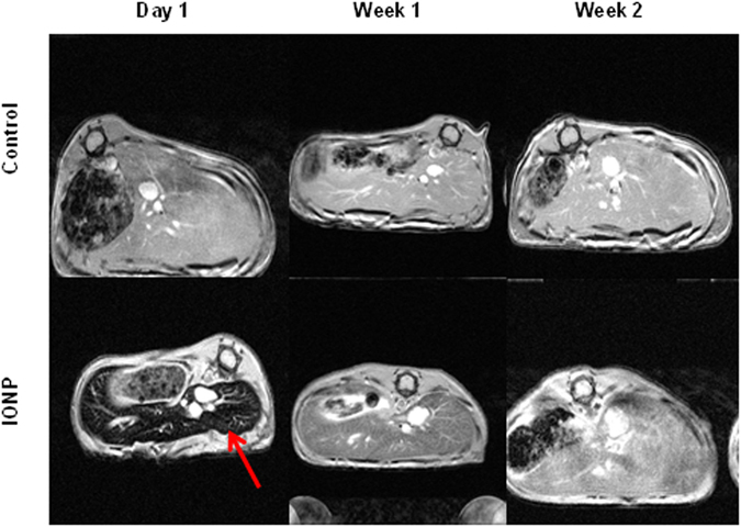
Stem cell tracking in cellular therapy and regenerative medicine is an urgent need, superparamagnetic iron oxide nanoparticles (IONPs) could be used as contrast agents in magnetic resonance imaging (MRI) that allows visualization of the implanted cells ensuring they reach the desired sites in vivo. Herein, we report the study of the interaction of 3,4-dihydroxyhydrocinnamic acid (DHCA) functionalized IONPs that have desirable properties for T - weighted MRI, with bone marrow-derived primary human mesenchymal stem cells (hMSCs). Using the multiparametric high-content imaging method, we evaluate cell viability, formation of reactive oxygen species, mitochondrial health, as well as cell morphology and determine that the hMSCs are minimally affected after labelling with IONPs. Their cellular uptake is visualized by transmission electron microscopy (TEM) and Prussian Blue staining, and quantified using an iron specific colourimetric method. In vitro and in vivo studies demonstrate that these IONPs are biocompatible and can produce significant contrast enhancement in T-weighted MRI. Iron oxide nanoparticles are detected in vivo as hypointense regions in the liver up to two weeks post injection using 9.4 T MRI. These DHCA functionalized IONPs are promising contrast agents for stem cell tracking by T-weighted MRI as they are biocompatible and show no evidence of cytotoxic effects on hMSCs.

摘要

细胞治疗和再生医学中的干细胞示踪是一项紧迫的需求,超顺磁性氧化铁纳米粒子(IONPs)可用作磁共振成像(MRI)中的对比剂,能够可视化植入的细胞,确保它们在体内到达预期的部位。在此,我们报告了 3,4-二羟基肉桂酸(DHCA)功能化 IONPs 与骨髓来源的原代人骨髓间充质干细胞(hMSCs)相互作用的研究。使用多参数高内涵成像方法,我们评估了细胞活力、活性氧的形成、线粒体健康以及细胞形态,并确定 hMSCs 在标记 IONPs 后受影响最小。通过透射电子显微镜(TEM)和普鲁士蓝染色观察到它们的细胞摄取,并使用铁特异性比色法进行定量。体外和体内研究表明,这些 IONPs 具有生物相容性,并可在 T 加权 MRI 中产生显著的对比增强。使用 9.4T MRI,在注射后两周内,这些 IONPs 在肝脏中作为低信号强度区域被体内检测到。这些 DHCA 功能化的 IONPs 是用于 T 加权 MRI 干细胞示踪的有前途的对比剂,因为它们具有生物相容性,并且对 hMSCs 没有显示出细胞毒性作用的证据。

https://cdn.ncbi.nlm.nih.gov/pmc/blobs/26f7/5552868/c932d9a45e7b/41598_2017_8092_Fig8_HTML.jpg
https://cdn.ncbi.nlm.nih.gov/pmc/blobs/26f7/5552868/86301ba50622/41598_2017_8092_Fig1_HTML.jpg
https://cdn.ncbi.nlm.nih.gov/pmc/blobs/26f7/5552868/f1ac4561d669/41598_2017_8092_Fig2_HTML.jpg
https://cdn.ncbi.nlm.nih.gov/pmc/blobs/26f7/5552868/68ee02f6f31e/41598_2017_8092_Fig3_HTML.jpg
https://cdn.ncbi.nlm.nih.gov/pmc/blobs/26f7/5552868/09fe32c90c6a/41598_2017_8092_Fig4_HTML.jpg
https://cdn.ncbi.nlm.nih.gov/pmc/blobs/26f7/5552868/4913df77b01b/41598_2017_8092_Fig5_HTML.jpg
https://cdn.ncbi.nlm.nih.gov/pmc/blobs/26f7/5552868/8b0defa34aad/41598_2017_8092_Fig6_HTML.jpg
https://cdn.ncbi.nlm.nih.gov/pmc/blobs/26f7/5552868/af5ee6d011c2/41598_2017_8092_Fig7_HTML.jpg
https://cdn.ncbi.nlm.nih.gov/pmc/blobs/26f7/5552868/c932d9a45e7b/41598_2017_8092_Fig8_HTML.jpg

相似文献

1
Assessing cell-nanoparticle interactions by high content imaging of biocompatible iron oxide nanoparticles as potential contrast agents for magnetic resonance imaging.通过高内涵成像评估生物相容性氧化铁纳米颗粒与细胞的相互作用,这些纳米颗粒可用作磁共振成像的潜在对比剂。
Sci Rep. 2017 Aug 10;7(1):7850. doi: 10.1038/s41598-017-08092-w.
2
Mesenchymal stromal cell labeling by new uncoated superparamagnetic maghemite nanoparticles in comparison with commercial Resovist--an initial in vitro study.新型未包被超顺磁性磁赤铁矿纳米颗粒与市售Resovist对比标记间充质基质细胞——一项初步体外研究
Int J Nanomedicine. 2014 Nov 20;9:5355-72. doi: 10.2147/IJN.S66986. eCollection 2014.
3
Synthesis Of PEG-Coated, Ultrasmall, Manganese-Doped Iron Oxide Nanoparticles With High Relaxivity For T/T Dual-Contrast Magnetic Resonance Imaging.聚乙二醇(PEG)包覆的超小粒径、高弛豫率锰掺杂氧化铁纳米粒子的合成及其在 T/T 双模态磁共振成像中的应用。
Int J Nanomedicine. 2019 Oct 24;14:8499-8507. doi: 10.2147/IJN.S219749. eCollection 2019.
4
Behaviour of adipose-derived canine mesenchymal stem cells after superparamagnetic iron oxide nanoparticles labelling for magnetic resonance imaging.超顺磁性氧化铁纳米颗粒标记后用于磁共振成像的犬脂肪来源间充质干细胞的行为
BMC Vet Res. 2017 Feb 24;13(1):62. doi: 10.1186/s12917-017-0980-0.
5
Magnetic resonance imaging of iron oxide labelled stem cells: applications to tissue engineering based regeneration of the intervertebral disc.氧化铁标记干细胞的磁共振成像:在基于组织工程的椎间盘再生中的应用
Eur Cell Mater. 2008 Aug 1;16:17-25. doi: 10.22203/ecm.v016a03.
6
Superparamagnetic iron oxide nanoparticles for direct labeling of stem cells and in vivo MRI tracking.用于干细胞直接标记和体内磁共振成像追踪的超顺磁性氧化铁纳米颗粒
Contrast Media Mol Imaging. 2016 Jan-Feb;11(1):55-64. doi: 10.1002/cmmi.1658. Epub 2015 Aug 3.
7
Efficient Labeling Of Mesenchymal Stem Cells For High Sensitivity Long-Term MRI Monitoring In Live Mice Brains.高效标记间充质干细胞用于活体小鼠大脑中高灵敏度的长期 MRI 监测。
Int J Nanomedicine. 2020 Jan 8;15:97-114. doi: 10.2147/IJN.S211205. eCollection 2020.
8
(Carboxymethyl)chitosan-modified superparamagnetic iron oxide nanoparticles for magnetic resonance imaging of stem cells.(羧甲基)壳聚糖修饰的超顺磁性氧化铁纳米颗粒用于干细胞的磁共振成像。
ACS Appl Mater Interfaces. 2009 Feb;1(2):328-35. doi: 10.1021/am8000538.
9
Design considerations for the synthesis of polymer coated iron oxide nanoparticles for stem cell labelling and tracking using MRI.用于使用 MRI 对干细胞进行标记和追踪的聚合物包覆氧化铁纳米粒子的合成设计考虑因素。
Chem Soc Rev. 2015 Oct 7;44(19):6733-48. doi: 10.1039/c5cs00331h.
10
Magnetic Resonance Imaging of Single Cells.磁共振成像技术在单细胞研究中的应用
Methods Mol Biol. 2020;2126:95-106. doi: 10.1007/978-1-0716-0364-2_9.

引用本文的文献

1
Applications and Efficacy of Iron Oxide Nanoparticles in the Treatment of Brain Tumors.氧化铁纳米颗粒在脑肿瘤治疗中的应用及疗效
Pharmaceutics. 2025 Apr 9;17(4):499. doi: 10.3390/pharmaceutics17040499.
2
Iron oxide based magnetic nanoparticles for hyperthermia, MRI and drug delivery applications: a review.用于热疗、磁共振成像和药物递送应用的氧化铁基磁性纳米颗粒:综述
RSC Adv. 2025 Apr 14;15(15):11587-11616. doi: 10.1039/d5ra00728c. eCollection 2025 Apr 9.
3
Fabrication of nanoparticles with precisely controllable plasmonic properties as tools for biomedical applications.

本文引用的文献

1
Controlled aggregation and cell uptake of thermoresponsive polyoxazoline-grafted superparamagnetic iron oxide nanoparticles.温敏性聚氧化烯接枝超顺磁性氧化铁纳米粒子的可控聚集和细胞摄取。
Nanoscale. 2017 Feb 23;9(8):2793-2805. doi: 10.1039/c6nr08654c.
2
Standardizing Nanomaterials.纳米材料标准化
ACS Nano. 2016 Nov 22;10(11):9763-9764. doi: 10.1021/acsnano.6b07629.
3
High throughput toxicity screening and intracellular detection of nanomaterials.纳米材料的高通量毒性筛选与细胞内检测
制备具有精确可控等离子体特性的纳米颗粒作为生物医学应用工具。
Nanoscale. 2025 Feb 20;17(8):4423-4438. doi: 10.1039/d4nr02677b.
4
Extraction of gamma iron oxide (γ-FeO) nanoparticles from waste can: Structure, morphology and magnetic properties.从废罐中提取γ-氧化铁(γ-FeO)纳米颗粒:结构、形态和磁性
Heliyon. 2024 May 9;10(10):e30810. doi: 10.1016/j.heliyon.2024.e30810. eCollection 2024 May 30.
5
Magnetic Hydroxyapatite Nanoparticles in Regenerative Medicine and Nanomedicine.再生医学与纳米医学中的磁性羟基磷灰石纳米颗粒
Int J Mol Sci. 2024 Feb 28;25(5):2809. doi: 10.3390/ijms25052809.
6
Functional Diversity in Radiolabeled Nanoceramics and Related Biomaterials for the Multimodal Imaging of Tumors.用于肿瘤多模态成像的放射性标记纳米陶瓷及相关生物材料的功能多样性
ACS Bio Med Chem Au. 2023 Aug 8;3(5):389-417. doi: 10.1021/acsbiomedchemau.3c00021. eCollection 2023 Oct 18.
7
Engineered Protein-Iron Oxide Hybrid Biomaterial for MRI-traceable Drug Encapsulation.用于MRI可追踪药物封装的工程化蛋白质-氧化铁杂化生物材料
Mol Syst Des Eng. 2022 Aug 1;7(8):915-932. doi: 10.1039/d2me00002d. Epub 2022 May 6.
8
Energetic Electron-Assisted Synthesis of Tailored Magnetite (FeO) and Maghemite (γ-FeO) Nanoparticles: Structure and Magnetic Properties.高能电子辅助合成定制磁铁矿(Fe₃O₄)和磁赤铁矿(γ-Fe₂O₃)纳米颗粒:结构与磁性
Nanomaterials (Basel). 2023 Feb 21;13(5):786. doi: 10.3390/nano13050786.
9
Nanotechnology strategies for hepatocellular carcinoma diagnosis and treatment.用于肝细胞癌诊断和治疗的纳米技术策略。
RSC Adv. 2022 Oct 31;12(48):31068-31082. doi: 10.1039/d2ra05127c. eCollection 2022 Oct 27.
10
Prodigious therapeutic effects of combining mesenchymal stem cells with magnetic nanoparticles.间充质干细胞与磁性纳米颗粒联合使用具有巨大的治疗效果。
World J Stem Cells. 2022 Jul 26;14(7):513-526. doi: 10.4252/wjsc.v14.i7.513.
Wiley Interdiscip Rev Nanomed Nanobiotechnol. 2017 Jan;9(1). doi: 10.1002/wnan.1413. Epub 2016 Jun 7.
4
Polyol synthesis, functionalisation, and biocompatibility studies of superparamagnetic iron oxide nanoparticles as potential MRI contrast agents.超顺磁性氧化铁纳米颗粒作为潜在磁共振成像造影剂的多元醇合成、功能化及生物相容性研究
Nanoscale. 2016 Feb 14;8(6):3278-87. doi: 10.1039/c5nr03867g. Epub 2015 Oct 13.
5
Biocompatible Low-Retention Superparamagnetic Iron Oxide Nanoclusters as Contrast Agents for Magnetic Resonance Imaging of Liver Tumor.生物相容性低保留超顺磁性氧化铁纳米团簇作为肝脏肿瘤磁共振成像的造影剂
J Biomed Nanotechnol. 2015 May;11(5):854-64. doi: 10.1166/jbn.2015.2042.
6
In vitro cytotoxicity of superparamagnetic iron oxide nanoparticles on neuronal and glial cells. Evaluation of nanoparticle interference with viability tests.超顺磁性氧化铁纳米颗粒对神经元和神经胶质细胞的体外细胞毒性。纳米颗粒对活力测试干扰的评估。
J Appl Toxicol. 2016 Mar;36(3):361-72. doi: 10.1002/jat.3213. Epub 2015 Jul 26.
7
Tissue-Engineered Tracheal Replacement in a Child: A 4-Year Follow-Up Study.儿童组织工程气管置换:一项4年随访研究
Am J Transplant. 2015 Oct;15(10):2750-7. doi: 10.1111/ajt.13318. Epub 2015 Jun 2.
8
Nanoparticle-based diagnostic imaging of inflammation in rheumatic disease.基于纳米颗粒的风湿性疾病炎症诊断成像
Curr Rheumatol Rev. 2014;10(1):3-10. doi: 10.2174/1573403x10666140914154816.
9
High-content imaging and gene expression analysis to study cell-nanomaterial interactions: the effect of surface hydrophobicity.高内涵成像和基因表达分析研究细胞-纳米材料相互作用:表面疏水性的影响。
Biomaterials. 2014 Dec;35(37):9941-9950. doi: 10.1016/j.biomaterials.2014.08.031. Epub 2014 Sep 15.
10
Widespread nanoparticle-assay interference: implications for nanotoxicity testing.广泛存在的纳米颗粒检测干扰:对纳米毒性测试的影响。
PLoS One. 2014 Mar 11;9(3):e90650. doi: 10.1371/journal.pone.0090650. eCollection 2014.