• 文献检索
  • 文档翻译
  • 深度研究
  • 学术资讯
  • Suppr Zotero 插件Zotero 插件
  • 邀请有礼
  • 套餐&价格
  • 历史记录
应用&插件
Suppr Zotero 插件Zotero 插件浏览器插件Mac 客户端Windows 客户端微信小程序
定价
高级版会员购买积分包购买API积分包
服务
文献检索文档翻译深度研究API 文档MCP 服务
关于我们
关于 Suppr公司介绍联系我们用户协议隐私条款
关注我们

Suppr 超能文献

核心技术专利:CN118964589B侵权必究
粤ICP备2023148730 号-1Suppr @ 2026

文献检索

告别复杂PubMed语法,用中文像聊天一样搜索,搜遍4000万医学文献。AI智能推荐,让科研检索更轻松。

立即免费搜索

文件翻译

保留排版,准确专业,支持PDF/Word/PPT等文件格式,支持 12+语言互译。

免费翻译文档

深度研究

AI帮你快速写综述,25分钟生成高质量综述,智能提取关键信息,辅助科研写作。

立即免费体验

用于肿瘤多模态成像的放射性标记纳米陶瓷及相关生物材料的功能多样性

Functional Diversity in Radiolabeled Nanoceramics and Related Biomaterials for the Multimodal Imaging of Tumors.

作者信息

Calatayud David G, Lledos Marina, Casarsa Federico, Pascu Sofia I

机构信息

Department of Inorganic Chemistry, Universidad Autónoma de Madrid, Madrid 28049, Spain.

Department of Electroceramics, Instituto de Cerámica y Vidrio, Madrid 28049, Spain.

出版信息

ACS Bio Med Chem Au. 2023 Aug 8;3(5):389-417. doi: 10.1021/acsbiomedchemau.3c00021. eCollection 2023 Oct 18.

DOI:10.1021/acsbiomedchemau.3c00021
PMID:37876497
原文链接:https://pmc.ncbi.nlm.nih.gov/articles/PMC10591303/
Abstract

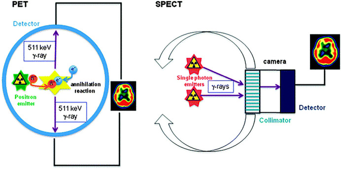
Nanotechnology advances have the potential to assist toward the earlier detection of diseases, giving increased accuracy for diagnosis and helping to personalize treatments, especially in the case of noncommunicative diseases (NCDs) such as cancer. The main advantage of nanoparticles, the scaffolds underpinning nanomedicine, is their potential to present multifunctionality: synthetic nanoplatforms for nanomedicines can be tailored to support a range of biomedical imaging modalities of relevance for clinical practice, such as, for example, optical imaging, computed tomography (CT), magnetic resonance imaging (MRI), single photon emission computed tomography (SPECT), and positron emission tomography (PET). A single nanoparticle has the potential to incorporate myriads of contrast agent units or imaging tracers, encapsulate, and/or be conjugated to different combinations of imaging tags, thus providing the means for multimodality diagnostic methods. These arrangements have been shown to provide significant improvements to the signal-to-noise ratios that may be obtained by molecular imaging techniques, for example, in PET diagnostic imaging with nanomaterials versus the cases when molecular species are involved as radiotracers. We surveyed some of the main discoveries in the simultaneous incorporation of nanoparticulate materials and imaging agents within highly kinetically stable radio-nanomaterials as potential tracers with (pre)clinical potential. Diversity in function and new developments toward synthesis, radiolabeling, and microscopy investigations are explored, and preclinical applications in molecular imaging are highlighted. The emphasis is on the biocompatible materials at the forefront of the main preclinical developments, e.g., nanoceramics and liposome-based constructs, which have driven the evolution of diagnostic radio-nanomedicines over the past decade.

摘要

纳米技术的进步有可能助力疾病的早期检测,提高诊断准确性并有助于实现个性化治疗,尤其是在癌症等非传染性疾病(NCDs)的情况下。纳米颗粒作为纳米医学的支撑框架,其主要优势在于具有呈现多功能性的潜力:用于纳米医学的合成纳米平台可以进行定制,以支持一系列与临床实践相关的生物医学成像模式,例如光学成像、计算机断层扫描(CT)、磁共振成像(MRI)、单光子发射计算机断层扫描(SPECT)和正电子发射断层扫描(PET)。单个纳米颗粒有可能包含无数的造影剂单元或成像示踪剂,封装和/或与不同组合的成像标签结合,从而为多模态诊断方法提供手段。这些配置已被证明能显著提高分子成像技术可能获得的信噪比,例如,在使用纳米材料的PET诊断成像中与分子物种作为放射性示踪剂的情况相比。我们调查了在高度动力学稳定的放射性纳米材料中同时掺入纳米颗粒材料和成像剂作为具有(临床前)潜力的潜在示踪剂的一些主要发现。探讨了功能的多样性以及合成、放射性标记和显微镜研究方面的新进展,并强调了分子成像中的临床前应用。重点是处于主要临床前发展前沿的生物相容性材料,例如纳米陶瓷和基于脂质体的构建体,它们在过去十年中推动了诊断性放射性纳米医学的发展。

https://cdn.ncbi.nlm.nih.gov/pmc/blobs/0a03/10591303/a8993da75adf/bg3c00021_0017.jpg
https://cdn.ncbi.nlm.nih.gov/pmc/blobs/0a03/10591303/499f8fbb70aa/bg3c00021_0001.jpg
https://cdn.ncbi.nlm.nih.gov/pmc/blobs/0a03/10591303/812a7efd8810/bg3c00021_0004.jpg
https://cdn.ncbi.nlm.nih.gov/pmc/blobs/0a03/10591303/d2ec09f581ab/bg3c00021_0002.jpg
https://cdn.ncbi.nlm.nih.gov/pmc/blobs/0a03/10591303/63d0085925a5/bg3c00021_0005.jpg
https://cdn.ncbi.nlm.nih.gov/pmc/blobs/0a03/10591303/404841a5d07f/bg3c00021_0006.jpg
https://cdn.ncbi.nlm.nih.gov/pmc/blobs/0a03/10591303/e366cf720251/bg3c00021_0007.jpg
https://cdn.ncbi.nlm.nih.gov/pmc/blobs/0a03/10591303/bd55d2de6bfa/bg3c00021_0008.jpg
https://cdn.ncbi.nlm.nih.gov/pmc/blobs/0a03/10591303/502983db7cad/bg3c00021_0009.jpg
https://cdn.ncbi.nlm.nih.gov/pmc/blobs/0a03/10591303/ba21067a2ad6/bg3c00021_0010.jpg
https://cdn.ncbi.nlm.nih.gov/pmc/blobs/0a03/10591303/770e27aa825d/bg3c00021_0011.jpg
https://cdn.ncbi.nlm.nih.gov/pmc/blobs/0a03/10591303/a78dd84b24c0/bg3c00021_0012.jpg
https://cdn.ncbi.nlm.nih.gov/pmc/blobs/0a03/10591303/16a275993c9f/bg3c00021_0013.jpg
https://cdn.ncbi.nlm.nih.gov/pmc/blobs/0a03/10591303/c0e37a29de24/bg3c00021_0014.jpg
https://cdn.ncbi.nlm.nih.gov/pmc/blobs/0a03/10591303/bcbf3e0b5e16/bg3c00021_0015.jpg
https://cdn.ncbi.nlm.nih.gov/pmc/blobs/0a03/10591303/33864419acdb/bg3c00021_0016.jpg
https://cdn.ncbi.nlm.nih.gov/pmc/blobs/0a03/10591303/a8993da75adf/bg3c00021_0017.jpg

相似文献

1
Functional Diversity in Radiolabeled Nanoceramics and Related Biomaterials for the Multimodal Imaging of Tumors.用于肿瘤多模态成像的放射性标记纳米陶瓷及相关生物材料的功能多样性
ACS Bio Med Chem Au. 2023 Aug 8;3(5):389-417. doi: 10.1021/acsbiomedchemau.3c00021. eCollection 2023 Oct 18.
2
Advanced Methods for Radiolabeling Multimodality Nanomedicines for SPECT/MRI and PET/MRI.用于 SPECT/MRI 和 PET/MRI 的多模态纳米医学放射性标记的先进方法。
J Nucl Med. 2018 Mar;59(3):382-389. doi: 10.2967/jnumed.116.187419. Epub 2017 Oct 12.
3
Exploring innovative strides in radiolabeled nanoparticle progress for multimodality cancer imaging and theranostic applications.探索放射性标记纳米颗粒在多模态癌症成像和治疗应用方面的创新进展。
Cancer Imaging. 2024 Sep 20;24(1):127. doi: 10.1186/s40644-024-00762-z.
4
Magnetic resonance imaging (MRI) for the assessment of myocardial viability: an evidence-based analysis.用于评估心肌存活性的磁共振成像(MRI):一项基于证据的分析。
Ont Health Technol Assess Ser. 2010;10(15):1-45. Epub 2010 Jul 1.
5
Positron emission tomography imaging using radiolabeled inorganic nanomaterials.使用放射性标记无机纳米材料的正电子发射断层扫描成像
Acc Chem Res. 2015 Feb 17;48(2):286-94. doi: 10.1021/ar500362y. Epub 2015 Jan 30.
6
Radiolabeling nanomaterials for multimodality imaging: New insights into nuclear medicine and cancer diagnosis.用于多模态成像的放射性标记纳米材料:核医学与癌症诊断的新见解
Biomaterials. 2020 Jan;228:119553. doi: 10.1016/j.biomaterials.2019.119553. Epub 2019 Oct 22.
7
An overview of nanoscale radionuclides and radiolabeled nanomaterials commonly used for nuclear molecular imaging and therapeutic functions.纳米尺度放射性核素和放射性标记纳米材料概述,通常用于核分子成像和治疗功能。
J Biomed Mater Res A. 2019 Jan;107(1):251-285. doi: 10.1002/jbm.a.36550. Epub 2018 Oct 25.
8
Nanoparticles and radiotracers: advances toward radionanomedicine.纳米颗粒与放射性示踪剂:放射性纳米医学的进展
Wiley Interdiscip Rev Nanomed Nanobiotechnol. 2016 Nov;8(6):872-890. doi: 10.1002/wnan.1402. Epub 2016 Mar 23.
9
Multimodality imaging in nanomedicine and nanotheranostics.纳米医学与纳米诊疗中的多模态成像
Cancer Biol Med. 2016 Sep;13(3):339-348. doi: 10.20892/j.issn.2095-3941.2016.0055.
10
Multimodality imaging: beyond PET/CT and SPECT/CT.多模态成像:超越PET/CT和SPECT/CT
Semin Nucl Med. 2009 Sep;39(5):348-53. doi: 10.1053/j.semnuclmed.2009.03.001.

引用本文的文献

1
Rare Earths-Doped and Ceria-Coated Strontium Aluminate PlateletsVersatile Luminescent Platforms for Correlated Lifetime Imaging by Multiphoton FLIM and PLIM.稀土掺杂及二氧化铈包覆的铝酸锶片状物——用于多光子荧光寿命成像(FLIM)和光致发光寿命成像(PLIM)相关寿命成像的多功能发光平台
ACS Omega. 2025 Apr 29;10(19):19950-19965. doi: 10.1021/acsomega.5c01649. eCollection 2025 May 20.
2
Protein-functionalized and intrinsically radiolabeled [Re]ReO nanoparticles: advancing cancer therapy through concurrent radio-photothermal effects.蛋白质功能化且具有固有放射性标记的[铼]ReO纳米颗粒:通过同步放射光热效应推进癌症治疗
Eur J Nucl Med Mol Imaging. 2025 Jan 25. doi: 10.1007/s00259-025-07074-9.

本文引用的文献

1
Nano-Theranostics for the Sensing, Imaging and Therapy of Prostate Cancers.用于前列腺癌传感、成像和治疗的纳米诊疗学
Front Chem. 2022 Apr 12;10:830133. doi: 10.3389/fchem.2022.830133. eCollection 2022.
2
Biocompatible Probes Based on Rare-Earth Doped Strontium Aluminates with Long-Lasting Phosphorescent Properties for In Vitro Optical IMAGING.基于长余辉发光特性的掺稀土 SrAl2O4 的生物相容性探针用于体外光学成像。
Int J Mol Sci. 2022 Mar 21;23(6):3410. doi: 10.3390/ijms23063410.
3
Review on Natural, Incidental, Bioinspired, and Engineered Nanomaterials: History, Definitions, Classifications, Synthesis, Properties, Market, Toxicities, Risks, and Regulations.
天然、偶然、仿生和工程纳米材料综述:历史、定义、分类、合成、性质、市场、毒性、风险及法规
Nanomaterials (Basel). 2022 Jan 6;12(2):177. doi: 10.3390/nano12020177.
4
Recent Advances in Nanomaterials Development for Nanomedicine and Cancer.纳米材料在纳米医学和癌症中的最新进展。
ACS Appl Bio Mater. 2021 Aug 16;4(8):5908-5925. doi: 10.1021/acsabm.1c00591. Epub 2021 Jul 19.
5
Liposome delivery to the brain with rapid short-pulses of focused ultrasound and microbubbles.脂质体经聚焦超声和微泡快速短脉冲递送至脑部。
J Control Release. 2022 Jan;341:605-615. doi: 10.1016/j.jconrel.2021.12.005. Epub 2021 Dec 10.
6
Nanoparticles for Cancer Therapy: Current Progress and Challenges.用于癌症治疗的纳米颗粒:当前进展与挑战
Nanoscale Res Lett. 2021 Dec 5;16(1):173. doi: 10.1186/s11671-021-03628-6.
7
Recent Progress in Technetium-99m-Labeled Nanoparticles for Molecular Imaging and Cancer Therapy.用于分子成像和癌症治疗的锝-99m标记纳米颗粒的最新进展
Nanomaterials (Basel). 2021 Nov 10;11(11):3022. doi: 10.3390/nano11113022.
8
Nanoparticles for Cancer Diagnosis, Radionuclide Therapy and Theranostics.用于癌症诊断、放射性核素治疗和治疗学的纳米粒子。
ACS Nano. 2021 Nov 23;15(11):16974-16981. doi: 10.1021/acsnano.1c09139. Epub 2021 Nov 8.
9
Metallodrugs are unique: opportunities and challenges of discovery and development.金属药物独具特色:发现与开发的机遇与挑战
Chem Sci. 2020 Nov 12;11(48):12888-12917. doi: 10.1039/d0sc04082g.
10
Hybrid Hierarchical Heterostructures of Nanoceramic Phosphors as Imaging Agents for Multiplexing and Living Cancer Cells Translocation.纳米陶瓷荧光粉的混合分层异质结构作为多重成像剂和活癌细胞迁移的示踪剂。
ACS Appl Bio Mater. 2021 May 17;4(5):4105-4118. doi: 10.1021/acsabm.0c01417. Epub 2021 Mar 10.