Wang Peng-Fei, Li Zhen-Guang, Zhang Yong, Ju Xiao-Hua, Liu Xin-Wu, Zhou Ai-Ming, Chen Jing
Department of Neurology, Weihai Municipal HospitalWeihai, China.
Department of Neurology, The Third Hospital of PLABaoji, China.
Front Cell Neurosci. 2017 Jul 14;11:206. doi: 10.3389/fncel.2017.00206. eCollection 2017.
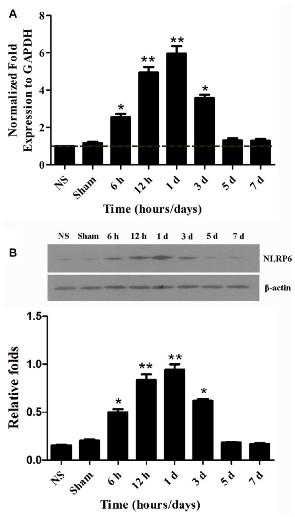
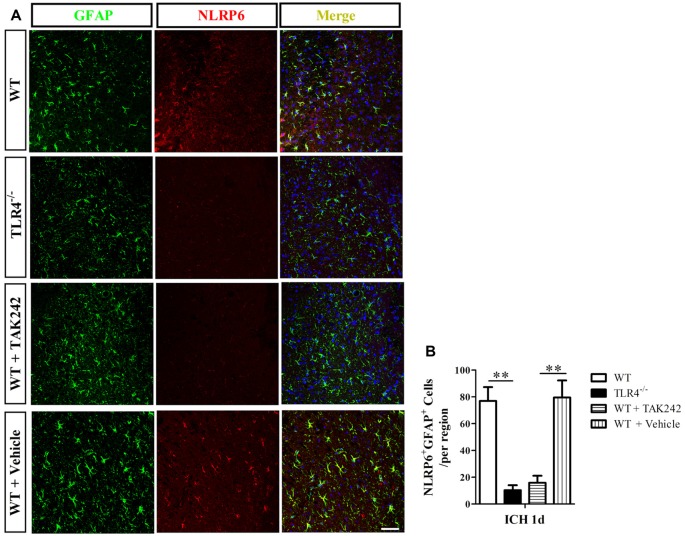
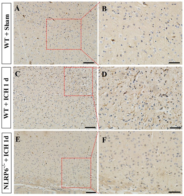
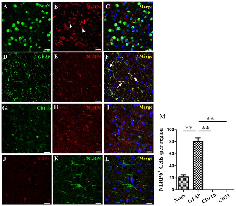
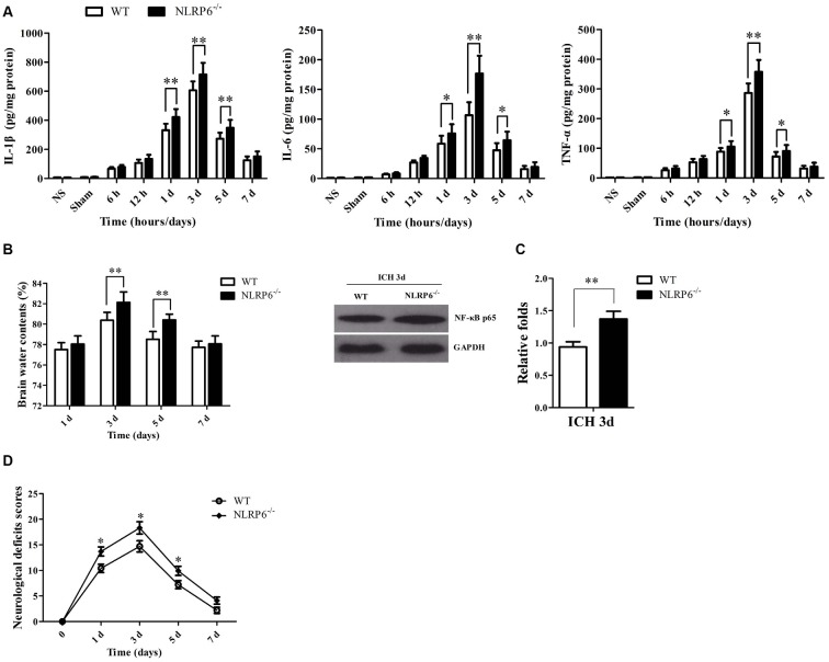
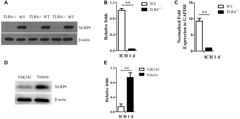
NLRP6 inflammasome, one of the important intracellular innate immune sensors, has been shown to regulate immune responses. However, its roles in the intracerebral hemorrhage (ICH) are completely not clear. In the present study, we investigated the expression profile and biological roles of NLRP6 inflammasome in perihematomal brain tissues of mice subjected to ICH. In this study, we investigated the expression profile of NLRP6 inflammasome in the perihematomal brain tissues and explored the biological role of NLRP6 inflammasome upon acute brain injury in mice subjected to ICH. Increased expression of NLRP6 inflammasome was found in perihematomal brain tissues ranging from 6 h to 3 days, with a peak level at 1 day after ICH. Immunohistochemistry staining also showed that NLRP6 inflammasome was significantly increased in the perihematomal brain tissues at 1 day after ICH. Moreover, immunofluorescence staining showed that NLRP6 inflammasome was mainly colocalized in glial fibrillary acidic protein (GFAP)-positive astrocytes, while with little colocalized expression in NeuN-positive neurons and without expression in CD11b-positive microglia and CD31-positive endothelial cell in the perihematomal brain tissue of mice after ICH. Furthermore, NLRP6 ICH mice exhibited significantly higher brain water contents (BMCs), proinflammatory cytokines, NF-κB activity and neurological deficit scores when compared with the wild type (WT) ICH mice. In addition, we found that Toll-like receptor 4 (TLR4) mice, as well as the TAK242 treated mice, had markedly lower expression of NLRP6 inflammasome expression in the perihematomal brain tissue at 1 day after ICH. Our data suggest that the upregulated NLRP6 inflammasome in perihematomal brain tissues attenuates ICH-induced brain injury.
NLRP6炎性小体是重要的细胞内固有免疫传感器之一,已被证明可调节免疫反应。然而,其在脑出血(ICH)中的作用尚完全不清楚。在本研究中,我们调查了脑出血小鼠血肿周围脑组织中NLRP6炎性小体的表达谱及其生物学作用。在本研究中,我们调查了血肿周围脑组织中NLRP6炎性小体的表达谱,并探讨了NLRP6炎性小体在脑出血小鼠急性脑损伤中的生物学作用。发现血肿周围脑组织中NLRP6炎性小体的表达在6小时至3天内增加,在脑出血后1天达到峰值水平。免疫组织化学染色也显示,脑出血后1天血肿周围脑组织中NLRP6炎性小体显著增加。此外,免疫荧光染色显示,NLRP6炎性小体主要与胶质纤维酸性蛋白(GFAP)阳性星形胶质细胞共定位,而在NeuN阳性神经元中几乎没有共定位表达,在脑出血后小鼠血肿周围脑组织的CD11b阳性小胶质细胞和CD31阳性内皮细胞中无表达。此外,与野生型(WT)脑出血小鼠相比,NLRP6脑出血小鼠的脑含水量(BMC)、促炎细胞因子、NF-κB活性和神经功能缺损评分显著更高。此外,我们发现Toll样受体4(TLR4)小鼠以及TAK242处理的小鼠在脑出血后1天血肿周围脑组织中NLRP6炎性小体的表达明显降低。我们的数据表明,血肿周围脑组织中上调的NLRP6炎性小体减轻了脑出血诱导的脑损伤。