Wang Yafei, Yu Dongsheng, Liu Zhiming, Zhou Fang, Dai Jun, Wu Bingbing, Zhou Jing, Heng Boon Chin, Zou Xiao Hui, Ouyang Hongwei, Liu Hua
Dr. Li Dak Sum & Yip Yio Chin Center for Stem Cell and Regenerative Medicine, Zhejiang University, Hangzhou, 310058, People's Republic of China.
Key Laboratory of Tissue Engineering and Regenerative Medicine of Zhejiang Province, Hangzhou, 310058, People's Republic of China.
Stem Cell Res Ther. 2017 Aug 14;8(1):189. doi: 10.1186/s13287-017-0632-0.
Mesenchymal stem cell therapy for osteoarthritis (OA) has been widely investigated, but the mechanisms are still unclear. Exosomes that serve as carriers of genetic information have been implicated in many diseases and are known to participate in many physiological processes. Here, we investigate the therapeutic potential of exosomes from human embryonic stem cell-induced mesenchymal stem cells (ESC-MSCs) in alleviating osteoarthritis (OA).
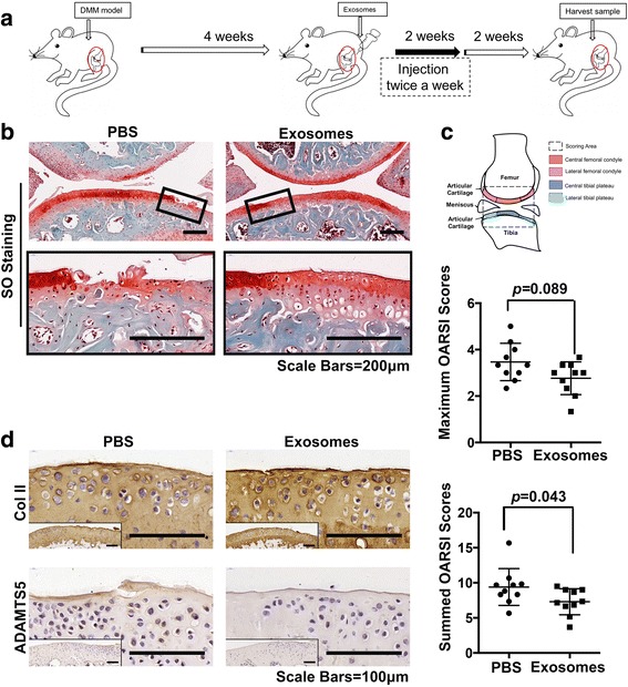
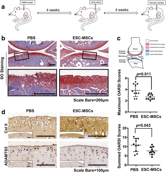
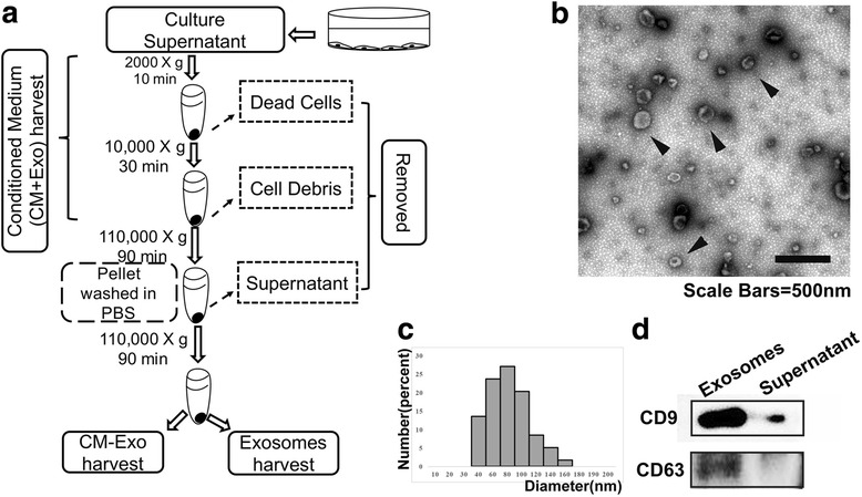
Exosomes were harvested from conditioned culture media of ESC-MSCs by a sequential centrifugation process. Primary mouse chondrocytes treated with interleukin 1 beta (IL-1β) were used as an in vitro model to evaluate the effects of the conditioned medium with or without exosomes and titrated doses of isolated exosomes for 48 hours, prior to immunocytochemistry or western blot analysis. Destabilization of the medial meniscus (DMM) surgery was performed on the knee joints of C57BL/6 J mice as an OA model. This was followed by intra-articular injection of either ESC-MSCs or their exosomes. Cartilage destruction and matrix degradation were evaluated with histological staining and OARSI scores at the post-surgery 8 weeks.
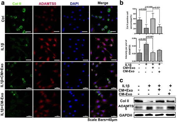
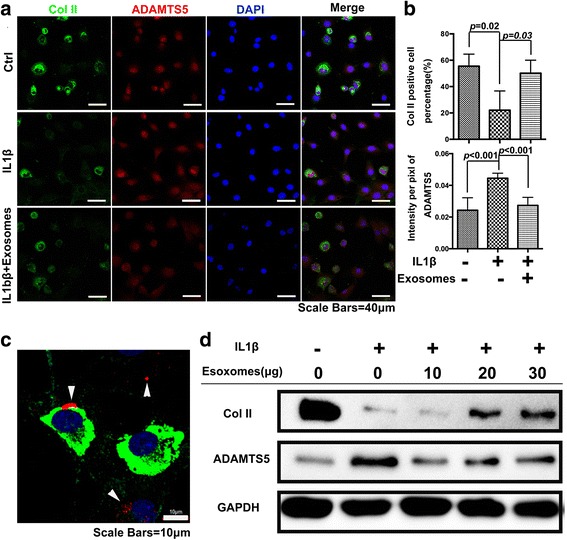
We found that intra-articular injection of ESC-MSCs alleviated cartilage destruction and matrix degradation in the DMM model. Further in vitro studies illustrated that this effect was exerted through ESC-MSC-derived exosomes. These exosomes maintained the chondrocyte phenotype by increasing collagen type II synthesis and decreasing ADAMTS5 expression in the presence of IL-1β. Immunocytochemistry revealed colocalization of the exosomes and collagen type II-positive chondrocytes. Subsequent intra-articular injection of exosomes derived from ESC-MSCs successfully impeded cartilage destruction in the DMM model.
The exosomes from ESC-MSCs exert a beneficial therapeutic effect on OA by balancing the synthesis and degradation of chondrocyte extracellular matrix (ECM), which in turn provides a new target for OA drug and drug-delivery system development.
间充质干细胞治疗骨关节炎(OA)已得到广泛研究,但其机制仍不清楚。作为遗传信息载体的外泌体与多种疾病有关,并且已知参与许多生理过程。在此,我们研究了人胚胎干细胞诱导的间充质干细胞(ESC-MSCs)来源的外泌体在缓解骨关节炎(OA)方面的治疗潜力。
通过连续离心法从ESC-MSCs的条件培养基中收获外泌体。用白细胞介素1β(IL-1β)处理的原代小鼠软骨细胞作为体外模型,在进行免疫细胞化学或蛋白质印迹分析之前,评估含或不含外泌体的条件培养基以及不同滴定剂量的分离外泌体作用48小时的效果。对C57BL/6 J小鼠的膝关节进行内侧半月板不稳定(DMM)手术作为OA模型。随后进行关节内注射ESC-MSCs或其外泌体。在术后8周用组织学染色和骨关节炎研究学会(OARSI)评分评估软骨破坏和基质降解情况。
我们发现关节内注射ESC-MSCs可减轻DMM模型中的软骨破坏和基质降解。进一步的体外研究表明,这种作用是通过ESC-MSCs来源的外泌体发挥的。在存在IL-1β的情况下,这些外泌体通过增加II型胶原蛋白合成和降低含血小板凝血酶敏感蛋白基的解聚素样金属蛋白酶5(ADAMTS5)表达来维持软骨细胞表型。免疫细胞化学显示外泌体与II型胶原蛋白阳性软骨细胞共定位。随后关节内注射ESC-MSCs来源的外泌体成功地阻止了DMM模型中的软骨破坏。
ESC-MSCs来源的外泌体通过平衡软骨细胞外基质(ECM)的合成与降解对OA发挥有益的治疗作用,这反过来为OA药物和药物递送系统的开发提供了新的靶点。