• 文献检索
  • 文档翻译
  • 深度研究
  • 学术资讯
  • Suppr Zotero 插件Zotero 插件
  • 邀请有礼
  • 套餐&价格
  • 历史记录
应用&插件
Suppr Zotero 插件Zotero 插件浏览器插件Mac 客户端Windows 客户端微信小程序
定价
高级版会员购买积分包购买API积分包
服务
文献检索文档翻译深度研究API 文档MCP 服务
关于我们
关于 Suppr公司介绍联系我们用户协议隐私条款
关注我们

Suppr 超能文献

核心技术专利:CN118964589B侵权必究
粤ICP备2023148730 号-1Suppr @ 2026

文献检索

告别复杂PubMed语法,用中文像聊天一样搜索,搜遍4000万医学文献。AI智能推荐,让科研检索更轻松。

立即免费搜索

文件翻译

保留排版,准确专业,支持PDF/Word/PPT等文件格式,支持 12+语言互译。

免费翻译文档

深度研究

AI帮你快速写综述,25分钟生成高质量综述,智能提取关键信息,辅助科研写作。

立即免费体验

二维剪切波弹性成像在甲状旁腺功能亢进患者甲状旁腺病变评估中的应用

2D-Shear Wave Elastography in the Evaluation of Parathyroid Lesions in Patients with Hyperparathyroidism.

作者信息

Golu Ioana, Sporea Ioan, Moleriu Lavinia, Tudor Anca, Cornianu Marioara, Vlad Adrian, Timar Romulus, Balas Melania, Amzar Daniela, Vlad Mihaela

机构信息

Department of Endocrinology, "Victor Babes" University of Medicine and Pharmacy, Timisoara, Romania.

Elastography Center, "Victor Babes" University of Medicine and Pharmacy, Timisoara, Romania.

出版信息

Int J Endocrinol. 2017;2017:9092120. doi: 10.1155/2017/9092120. Epub 2017 Aug 6.

DOI:10.1155/2017/9092120
PMID:28845158
原文链接:https://pmc.ncbi.nlm.nih.gov/articles/PMC5563409/
Abstract

BACKGROUND AND AIMS

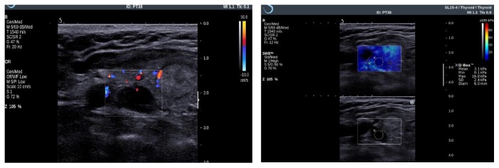
2D-shear wave elastography (2D-SWE) is a relatively new elastographic technique. The aim of the present study is to determine the values of the elasticity indexes (EI) measured by 2D-SWE in parathyroid benign lesions (adenomas or hyperplasia) and to establish if this investigation is helpful for the preoperative identification of the parathyroid adenoma.

MATERIAL AND METHODS

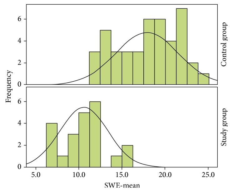
The study groups were represented by 22 patients with primary or tertiary hyperparathyroidism, diagnosed by specific tests, and 43 healthy controls, in whom the thyroid parenchyma was evaluated, in order to compare the EI of the thyroid tissue with those of the parathyroid lesions.

RESULTS

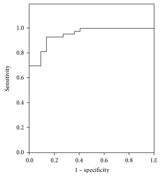
The mean EI measured by 2D-SWE in the parathyroid lesions was 10.2 ± 4.9 kPa, significantly lower than that of the normal thyroid parenchyma (19.5 ± 7.6 kPa; = 0.007), indicating soft tissue. For a cutoff value of 12.5 kPa, the EI assessed by 2D-SWE had a sensitivity of 93% and a specificity of 86% (AUC = 0.949; < 0.001) for predicting parathyroid lesions.

CONCLUSION

A value lower than 12.5 kPa for the mean EI measured by 2D-SWE can be used to confirm that the lesion/nodule is a parathyroid adenoma.

摘要

背景与目的

二维剪切波弹性成像(2D-SWE)是一种相对较新的弹性成像技术。本研究的目的是确定通过二维剪切波弹性成像测量的弹性指数(EI)在甲状旁腺良性病变(腺瘤或增生)中的值,并确定该检查是否有助于甲状旁腺腺瘤的术前识别。

材料与方法

研究组包括22例经特定检查诊断为原发性或继发性甲状旁腺功能亢进的患者以及43例健康对照者,对他们的甲状腺实质进行评估,以便比较甲状腺组织与甲状旁腺病变的弹性指数。

结果

二维剪切波弹性成像测量的甲状旁腺病变的平均弹性指数为10.2±4.9kPa,显著低于正常甲状腺实质(19.5±7.6kPa;P = 0.007),表明为软组织。对于12.5kPa的临界值,二维剪切波弹性成像评估的弹性指数对甲状旁腺病变的预测敏感性为93%,特异性为86%(AUC = 0.949;P < 0.001)。

结论

二维剪切波弹性成像测量的平均弹性指数低于12.5kPa的值可用于确认病变/结节为甲状旁腺腺瘤。

https://cdn.ncbi.nlm.nih.gov/pmc/blobs/4237/5563409/224396c00b7e/IJE2017-9092120.003.jpg
https://cdn.ncbi.nlm.nih.gov/pmc/blobs/4237/5563409/1eed927d6e2b/IJE2017-9092120.001.jpg
https://cdn.ncbi.nlm.nih.gov/pmc/blobs/4237/5563409/63e5f25aee8e/IJE2017-9092120.002.jpg
https://cdn.ncbi.nlm.nih.gov/pmc/blobs/4237/5563409/224396c00b7e/IJE2017-9092120.003.jpg
https://cdn.ncbi.nlm.nih.gov/pmc/blobs/4237/5563409/1eed927d6e2b/IJE2017-9092120.001.jpg
https://cdn.ncbi.nlm.nih.gov/pmc/blobs/4237/5563409/63e5f25aee8e/IJE2017-9092120.002.jpg
https://cdn.ncbi.nlm.nih.gov/pmc/blobs/4237/5563409/224396c00b7e/IJE2017-9092120.003.jpg

相似文献

1
2D-Shear Wave Elastography in the Evaluation of Parathyroid Lesions in Patients with Hyperparathyroidism.二维剪切波弹性成像在甲状旁腺功能亢进患者甲状旁腺病变评估中的应用
Int J Endocrinol. 2017;2017:9092120. doi: 10.1155/2017/9092120. Epub 2017 Aug 6.
2
Shear Wave Elastography versus Strain Elastography in Diagnosing Parathyroid Adenomas.剪切波弹性成像与应变弹性成像在诊断甲状旁腺腺瘤中的应用比较
Int J Endocrinol. 2020 Mar 17;2020:3801902. doi: 10.1155/2020/3801902. eCollection 2020.
3
Shear Wave Elastography in Patients with Primary and Secondary Hyperparathyroidism.原发性和继发性甲状旁腺功能亢进患者的剪切波弹性成像
J Clin Med. 2021 Feb 10;10(4):697. doi: 10.3390/jcm10040697.
4
Shear Wave Elastography in Diagnosing Secondary Hyperparathyroidism.剪切波弹性成像在诊断继发性甲状旁腺功能亢进中的应用
Diagnostics (Basel). 2019 Dec 5;9(4):213. doi: 10.3390/diagnostics9040213.
5
Differentiating thyroid nodules parathyroid lesions using 2D-shear-wave elastography: a novel approach for enhanced diagnostic accuracy.应用二维剪切波弹性成像技术鉴别甲状腺结节与甲状旁腺病变:提高诊断准确性的新方法。
Front Endocrinol (Lausanne). 2023 Jul 31;14:1231784. doi: 10.3389/fendo.2023.1231784. eCollection 2023.
6
Shear wave elastography for differentiating parathyroid neoplasms with malignant diagnosis or uncertain malignant potential from parathyroid adenomas: initial experience.超声剪切波弹性成像鉴别甲状旁腺良恶性肿瘤或性质不明的肿瘤与甲状旁腺腺瘤:初步经验。
Cancer Imaging. 2022 Nov 22;22(1):64. doi: 10.1186/s40644-022-00503-0.
7
Shear wave elastography in the diagnostics of parathyroid adenomas-new application of the method.剪切波弹性成像在甲状旁腺腺瘤诊断中的应用——方法的新应用。
Endocrine. 2018 May;60(2):240-245. doi: 10.1007/s12020-018-1553-0. Epub 2018 Feb 21.
8
Three-Dimensional Shear Wave Elastography for Differentiating Benign From Malignant Thyroid Nodules.用于鉴别甲状腺良恶性结节的三维剪切波弹性成像
J Ultrasound Med. 2018 Jul;37(7):1777-1788. doi: 10.1002/jum.14531. Epub 2018 Jan 9.
9
Evaluation of Parathyroid Lesions With Point Shear Wave Elastography.利用点剪切波弹性成像技术评估甲状旁腺病变
J Ultrasound Med. 2016 Oct;35(10):2179-82. doi: 10.7863/ultra.15.10074. Epub 2016 Aug 29.
10
Reappraisal of shear wave elastography as a diagnostic tool for identifying thyroid carcinoma.重新评估剪切波弹性成像作为诊断甲状腺癌的工具。
Endocr Connect. 2019 Aug;8(8):1195-1205. doi: 10.1530/EC-19-0324.

引用本文的文献

1
Shear wave elastography for differentiating parathyroid neoplasms with malignant diagnosis or uncertain malignant potential from parathyroid adenomas: initial experience.超声剪切波弹性成像鉴别甲状旁腺良恶性肿瘤或性质不明的肿瘤与甲状旁腺腺瘤:初步经验。
Cancer Imaging. 2022 Nov 22;22(1):64. doi: 10.1186/s40644-022-00503-0.
2
Pediatric hyperparathyroidism: review and imaging update.儿童甲状旁腺功能亢进症:综述与影像学进展。
Pediatr Radiol. 2021 Jun;51(7):1106-1120. doi: 10.1007/s00247-021-05050-7. Epub 2021 Apr 27.
3
Two-Dimensional Shear Wave Elastography of Normal Soft Tissue Organs in Adult Beagle Dogs; Interobserver Agreement and Sources of Variability.

本文引用的文献

1
Can ARFI elastography be used to differentiate parathyroid from thyroid lesions?声辐射力脉冲成像技术能否用于鉴别甲状旁腺与甲状腺病变?
J Endocrinol Invest. 2018 Jan;41(1):111-119. doi: 10.1007/s40618-017-0694-y. Epub 2017 May 31.
2
Evaluation of Parathyroid Lesions With Point Shear Wave Elastography.利用点剪切波弹性成像技术评估甲状旁腺病变
J Ultrasound Med. 2016 Oct;35(10):2179-82. doi: 10.7863/ultra.15.10074. Epub 2016 Aug 29.
3
The American Association of Endocrine Surgeons Guidelines for Definitive Management of Primary Hyperparathyroidism.
成年比格犬正常软组织器官的二维剪切波弹性成像:观察者间一致性及变异来源
Front Bioeng Biotechnol. 2020 Aug 19;8:979. doi: 10.3389/fbioe.2020.00979. eCollection 2020.
4
Preoperative and Intraoperative Methods of Parathyroid Gland Localization and the Diagnosis of Parathyroid Adenomas.甲状旁腺定位的术前和术中方法及甲状旁腺瘤的诊断。
Molecules. 2020 Apr 9;25(7):1724. doi: 10.3390/molecules25071724.
5
Ultrasound elastography score and strain index in different parathyroid lesions.不同甲状旁腺病变中的超声弹性成像评分及应变指数
Endocr Connect. 2019 Dec;8(12):1579-1590. doi: 10.1530/EC-19-0443.
6
Diagnostic Performance of Multiple Sound Touch Elastography for Differentiating Benign and Malignant Thyroid Nodules.多种声触诊弹性成像技术鉴别甲状腺良恶性结节的诊断性能
Front Pharmacol. 2018 Nov 26;9:1359. doi: 10.3389/fphar.2018.01359. eCollection 2018.
7
Shear wave elastography in the diagnostics of parathyroid adenomas-new application of the method.剪切波弹性成像在甲状旁腺腺瘤诊断中的应用——方法的新应用。
Endocrine. 2018 May;60(2):240-245. doi: 10.1007/s12020-018-1553-0. Epub 2018 Feb 21.
美国内分泌外科学会原发性甲状旁腺功能亢进症确定性治疗指南。
JAMA Surg. 2016 Oct 1;151(10):959-968. doi: 10.1001/jamasurg.2016.2310.
4
Shear wave elastography and parathyroid adenoma: A new tool for diagnosing parathyroid adenomas.剪切波弹性成像与甲状旁腺腺瘤:一种诊断甲状旁腺腺瘤的新工具。
Eur J Radiol. 2016 Sep;85(9):1586-93. doi: 10.1016/j.ejrad.2016.06.009. Epub 2016 Jun 17.
5
Ultrasound Elastography for Distinction Between Parathyroid Adenomas and Thyroid Nodules.超声弹性成像用于鉴别甲状旁腺腺瘤与甲状腺结节
J Ultrasound Med. 2016 Jun;35(6):1277-82. doi: 10.7863/ultra.15.07043. Epub 2016 May 5.
6
The role of ultrasound and nuclear medicine methods in the preoperative diagnostics of primary hyperparathyroidism.超声和核医学方法在原发性甲状旁腺功能亢进症术前诊断中的作用。
J Ultrason. 2015 Dec;15(63):398-409. doi: 10.15557/JoU.2015.0037. Epub 2015 Dec 28.
7
Shear Wave Elastography in Head and Neck Lymph Node Assessment: Image Quality and Diagnostic Impact Compared with B-Mode and Doppler Ultrasonography.剪切波弹性成像在头颈部淋巴结评估中的应用:与B型超声和多普勒超声相比的图像质量及诊断影响
Ultrasound Med Biol. 2016 Feb;42(2):387-98. doi: 10.1016/j.ultrasmedbio.2015.10.019. Epub 2015 Nov 23.
8
Virtual Touch Tissue Imaging Quantification Shear Wave Elastography: Prospective Assessment of Cervical Lymph Nodes.虚拟触诊组织成像定量剪切波弹性成像:对颈部淋巴结的前瞻性评估
Ultrasound Med Biol. 2016 Feb;42(2):378-86. doi: 10.1016/j.ultrasmedbio.2015.10.003. Epub 2015 Nov 6.
9
Real-time shear wave elastography may predict autoimmune thyroid disease.实时剪切波弹性成像可能预测自身免疫性甲状腺疾病。
Wien Klin Wochenschr. 2015 May;127(9-10):330-6. doi: 10.1007/s00508-015-0754-2. Epub 2015 Apr 3.
10
WFUMB guidelines and recommendations for clinical use of ultrasound elastography: Part 1: basic principles and terminology.世界超声医学与生物学联合会超声弹性成像临床应用指南与建议:第1部分:基本原则与术语
Ultrasound Med Biol. 2015 May;41(5):1126-47. doi: 10.1016/j.ultrasmedbio.2015.03.009. Epub 2015 Mar 21.