• 文献检索
  • 文档翻译
  • 深度研究
  • 学术资讯
  • Suppr Zotero 插件Zotero 插件
  • 邀请有礼
  • 套餐&价格
  • 历史记录
应用&插件
Suppr Zotero 插件Zotero 插件浏览器插件Mac 客户端Windows 客户端微信小程序
定价
高级版会员购买积分包购买API积分包
服务
文献检索文档翻译深度研究API 文档MCP 服务
关于我们
关于 Suppr公司介绍联系我们用户协议隐私条款
关注我们

Suppr 超能文献

核心技术专利:CN118964589B侵权必究
粤ICP备2023148730 号-1Suppr @ 2026

文献检索

告别复杂PubMed语法,用中文像聊天一样搜索,搜遍4000万医学文献。AI智能推荐,让科研检索更轻松。

立即免费搜索

文件翻译

保留排版,准确专业,支持PDF/Word/PPT等文件格式,支持 12+语言互译。

免费翻译文档

深度研究

AI帮你快速写综述,25分钟生成高质量综述,智能提取关键信息,辅助科研写作。

立即免费体验

重新评估剪切波弹性成像作为诊断甲状腺癌的工具。

Reappraisal of shear wave elastography as a diagnostic tool for identifying thyroid carcinoma.

作者信息

Swan Kristine Zøylner, Bonnema Steen Joop, Jespersen Marie Louise, Nielsen Viveque Egsgaard

机构信息

Department of Otorhinolaryngology Head & Neck Surgery, Aarhus University Hospital, Aarhus N, Denmark.

Department of Clinical Medicine, Health, Aarhus University, Aarhus N, Denmark.

出版信息

Endocr Connect. 2019 Aug;8(8):1195-1205. doi: 10.1530/EC-19-0324.

DOI:10.1530/EC-19-0324
PMID:31340198
原文链接:https://pmc.ncbi.nlm.nih.gov/articles/PMC6709542/
Abstract

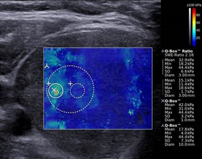
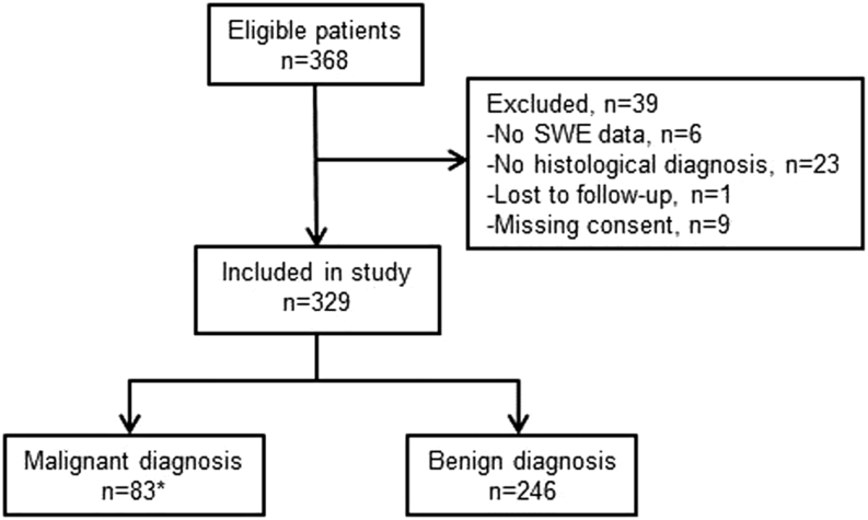
Thyroid nodular disease is common, but predicting the risk of malignancy can be difficult. In this prospective study, we aimed to assess the diagnostic accuracy of shear wave elastography (SWE) in predicting thyroid malignancy. Patients with thyroid nodules were enrolled from a surgical tertiary unit. Elasticity index (EI) measured by SWE was registered for seven EI outcomes assessing nodular stiffness and heterogeneity. The diagnosis was determined histologically. In total, 329 patients (mean age: 55 ± 13 years) with 413 thyroid nodules (mean size: 32 ± 13 mm, 88 malignant) were enrolled. Values of SWE region of interest (ROI) for malignant and benign nodules were highly overlapping (ranges for SWE-ROImean: malignant 3-100 kPa; benign 4-182 kPa), and no difference between malignant and benign nodules was found for any other EI outcome investigated (P = 0.13-0.96). There was no association between EI and the histological diagnosis by receiver operating characteristics analysis (area under the curve: 0.51-0.56). Consequently, defining a cut-off point of EI for the prediction of malignancy was not clinically meaningful. Testing our data on previously proposed cut-off points revealed a low accuracy of SWE (56-80%). By regression analysis, factors affecting EI included nodule size >30 mm, heterogeneous echogenicity, micro- or macrocalcifications and solitary nodule. In conclusion, EI, measured by SWE, showed huge overlap between malignant and benign nodules, and low diagnostic accuracy in the prediction of thyroid malignancy. Our study supports that firmness of thyroid nodules, as assessed by SWE, should not be a key feature in the evaluation of such lesions.

摘要

甲状腺结节性疾病很常见,但预测其恶性风险可能具有挑战性。在这项前瞻性研究中,我们旨在评估剪切波弹性成像(SWE)预测甲状腺恶性肿瘤的诊断准确性。甲状腺结节患者来自一家外科三级医疗单位。通过SWE测量的弹性指数(EI)被记录为七个评估结节硬度和异质性的EI结果。诊断通过组织学确定。总共纳入了329例患者(平均年龄:55±13岁),有413个甲状腺结节(平均大小:32±13mm,88个为恶性)。恶性和良性结节的SWE感兴趣区域(ROI)值高度重叠(SWE-ROImean范围:恶性为3-100kPa;良性为4-182kPa),并且在所研究的任何其他EI结果中,恶性和良性结节之间均未发现差异(P = 0.13-0.96)。通过受试者工作特征分析,EI与组织学诊断之间无关联(曲线下面积:0.51-0.56)。因此,定义EI的临界值以预测恶性肿瘤在临床上并无意义。用先前提出的临界值测试我们的数据显示SWE的准确性较低(56-80%)。通过回归分析,影响EI的因素包括结节大小>30mm、回声不均匀、微钙化或大钙化以及孤立结节。总之,通过SWE测量的EI显示恶性和良性结节之间有很大重叠,且在预测甲状腺恶性肿瘤方面诊断准确性较低。我们的研究支持,通过SWE评估的甲状腺结节硬度不应成为评估此类病变的关键特征。

https://cdn.ncbi.nlm.nih.gov/pmc/blobs/6f7b/6709542/6b5184910a2a/EC-19-0324fig2.jpg
https://cdn.ncbi.nlm.nih.gov/pmc/blobs/6f7b/6709542/8fcfa8cbe031/EC-19-0324fig1.jpg
https://cdn.ncbi.nlm.nih.gov/pmc/blobs/6f7b/6709542/6b5184910a2a/EC-19-0324fig2.jpg
https://cdn.ncbi.nlm.nih.gov/pmc/blobs/6f7b/6709542/8fcfa8cbe031/EC-19-0324fig1.jpg
https://cdn.ncbi.nlm.nih.gov/pmc/blobs/6f7b/6709542/6b5184910a2a/EC-19-0324fig2.jpg

相似文献

1
Reappraisal of shear wave elastography as a diagnostic tool for identifying thyroid carcinoma.重新评估剪切波弹性成像作为诊断甲状腺癌的工具。
Endocr Connect. 2019 Aug;8(8):1195-1205. doi: 10.1530/EC-19-0324.
2
The Role of Shear Wave Elastography in the Discrimination Between Malignant and Benign Thyroid Nodules.剪切波弹性成像在鉴别甲状腺良恶性结节中的作用
Acta Inform Med. 2020 Dec;28(4):248-253. doi: 10.5455/aim.2020.28.248-253.
3
Shear wave elasticity by tracing total nodule showed high reproducibility and concordance with fibrosis in thyroid cancer.通过追踪整个结节的剪切波弹性,发现甲状腺癌的弹性具有很高的可重复性和与纤维化的一致性。
BMC Cancer. 2020 Feb 12;20(1):118. doi: 10.1186/s12885-019-6437-z.
4
Three-Dimensional Shear Wave Elastography for Differentiating Benign From Malignant Thyroid Nodules.用于鉴别甲状腺良恶性结节的三维剪切波弹性成像
J Ultrasound Med. 2018 Jul;37(7):1777-1788. doi: 10.1002/jum.14531. Epub 2018 Jan 9.
5
Evaluation of thyroid micro-carcinoma using shear wave elastography: Initial experience with qualitative and quantitative analysis.应用剪切波弹性成像技术评估甲状腺微小癌:定性和定量分析的初步经验。
Eur J Radiol. 2021 Apr;137:109571. doi: 10.1016/j.ejrad.2021.109571. Epub 2021 Jan 29.
6
Diagnostic Performance of Shear Wave Elastography Parameters Alone and in Combination with Conventional B-Mode Ultrasound Parameters for the Characterization of Thyroid Nodules: A Prospective, Dual-Center Study.单独及联合传统B超参数的剪切波弹性成像参数对甲状腺结节特征的诊断性能:一项前瞻性双中心研究
Ultrasound Med Biol. 2016 Dec;42(12):2803-2811. doi: 10.1016/j.ultrasmedbio.2016.07.010. Epub 2016 Sep 9.
7
Shear wave elastography of thyroid nodules for the prediction of malignancy in a large scale study.在一项大规模研究中,甲状腺结节的剪切波弹性成像用于预测恶性肿瘤。
Eur J Radiol. 2015 Mar;84(3):407-412. doi: 10.1016/j.ejrad.2014.11.019. Epub 2014 Nov 27.
8
Diagnostic Performance of a Combination of Shear Wave Elastography and B-Mode Ultrasonography in Differentiating Benign From Malignant Thyroid Nodules.剪切波弹性成像与B型超声联合应用对甲状腺良恶性结节的诊断效能
Clin Exp Otorhinolaryngol. 2020 May;13(2):186-193. doi: 10.21053/ceo.2019.01235. Epub 2020 Mar 11.
9
Shear wave elastography of thyroid nodules in routine clinical practice: preliminary observations and utility for detecting malignancy.常规临床实践中的甲状腺结节剪切波弹性成像:初步观察及其在检测恶性肿瘤中的应用。
Eur Radiol. 2012 Nov;22(11):2397-406. doi: 10.1007/s00330-012-2495-1. Epub 2012 May 30.
10
Comparison of strain elastography, point shear wave elastography using acoustic radiation force impulse imaging and 2D-shear wave elastography for the differentiation of thyroid nodules.应变弹性成像、声辐射力脉冲成像点剪切波弹性成像与二维剪切波弹性成像在甲状腺结节鉴别诊断中的比较。
PLoS One. 2018 Sep 17;13(9):e0204095. doi: 10.1371/journal.pone.0204095. eCollection 2018.

引用本文的文献

1
Exploring the Potential of ChatGPT-4o in Thyroid Nodule Diagnosis Using Multi-Modality Ultrasound Imaging: Dual- vs. Triple-Modality Approaches.利用多模态超声成像探索ChatGPT-4o在甲状腺结节诊断中的潜力:双模态与三模态方法
Cancers (Basel). 2025 Jun 20;17(13):2068. doi: 10.3390/cancers17132068.
2
Diagnostic performance of shear wave elastography in thyroid nodules with indeterminate cytology: A systematic review and meta-analysis.剪切波弹性成像在甲状腺结节细针穿刺结果不确定中的诊断效能:一项系统评价和荟萃分析
Heliyon. 2023 Oct 5;9(10):e20654. doi: 10.1016/j.heliyon.2023.e20654. eCollection 2023 Oct.
3
Combined Shear Wave Elastography and EU TIRADS in Differentiating Malignant and Benign Thyroid Nodules.

本文引用的文献

1
Is shear wave elastography relevant in the diagnosis of polycystic ovarian syndrome?剪切波弹性成像在多囊卵巢综合征的诊断中是否具有相关性?
Med Ultrason. 2019 May 2;21(2):158-162. doi: 10.11152/mu-1849.
2
A multicentre validation study for the EU-TIRADS using histological diagnosis as a gold standard.一项以组织学诊断为金标准的针对 EU-TIRADS 的多中心验证研究。
Clin Endocrinol (Oxf). 2019 Aug;91(2):340-347. doi: 10.1111/cen.13997. Epub 2019 May 3.
3
European Thyroid Association Guidelines for Ultrasound Malignancy Risk Stratification of Thyroid Nodules in Adults: The EU-TIRADS.
联合剪切波弹性成像与欧盟甲状腺影像报告和数据系统在鉴别甲状腺良恶性结节中的应用
Cancers (Basel). 2022 Nov 10;14(22):5521. doi: 10.3390/cancers14225521.
4
Added value of mass characteristic frequency to 2-D shear wave elastography for differentiation of benign and malignant thyroid nodules.二维剪切波弹性成像中质量特征频率对良恶性甲状腺结节的鉴别诊断价值。
Ultrasound Med Biol. 2022 Aug;48(8):1663-1671. doi: 10.1016/j.ultrasmedbio.2022.04.218. Epub 2022 Jun 4.
5
External validation of AIBx, an artificial intelligence model for risk stratification, in thyroid nodules.用于甲状腺结节风险分层的人工智能模型AIBx的外部验证
Eur Thyroid J. 2022 Mar 8;11(2):e210129. doi: 10.1530/ETJ-21-0129.
6
Correlation between elastic modulus and clinical severity of pathological scars: a cross-sectional study.弹性模量与病理性瘢痕临床严重程度的相关性:一项横断面研究。
Sci Rep. 2021 Dec 2;11(1):23324. doi: 10.1038/s41598-021-02730-0.
7
Clinical Value of Shear Wave Elastography Color Scores in Classifying Thyroid Nodules.剪切波弹性成像彩色评分在甲状腺结节分类中的临床价值
Int J Gen Med. 2021 Nov 10;14:8007-8018. doi: 10.2147/IJGM.S331406. eCollection 2021.
8
Evaluation of thyroid nodules by shear wave elastography: a review of current knowledge.剪切波弹性成像评估甲状腺结节:当前知识综述。
J Endocrinol Invest. 2021 Oct;44(10):2043-2056. doi: 10.1007/s40618-021-01570-z. Epub 2021 Apr 16.
9
The Role of Shear Wave Elastography in the Discrimination Between Malignant and Benign Thyroid Nodules.剪切波弹性成像在鉴别甲状腺良恶性结节中的作用
Acta Inform Med. 2020 Dec;28(4):248-253. doi: 10.5455/aim.2020.28.248-253.
10
Multimode ultrasonic technique is recommended for the differential diagnosis of thyroid cancer.多模式超声技术被推荐用于甲状腺癌的鉴别诊断。
PeerJ. 2020 May 4;8:e9112. doi: 10.7717/peerj.9112. eCollection 2020.
欧洲甲状腺协会成人甲状腺结节超声恶性风险分层指南:欧盟甲状腺影像报告和数据系统(EU-TIRADS)
Eur Thyroid J. 2017 Sep;6(5):225-237. doi: 10.1159/000478927. Epub 2017 Aug 8.
4
Is Diagnostic Performance of Quantitative 2D-Shear Wave Elastography Optimal for Clinical Classification of Benign and Malignant Thyroid Nodules?: A Systematic Review and Meta-analysis.定量 2D 剪切波弹性成像的诊断性能是否最适合甲状腺良恶性结节的临床分类?:系统评价和荟萃分析。
Acad Radiol. 2022 Mar;29 Suppl 3:S114-S121. doi: 10.1016/j.acra.2017.09.002. Epub 2017 Oct 18.
5
Value of shear wave elastography in discriminating malignant and benign breast lesions: A meta-analysis.剪切波弹性成像在鉴别乳腺良恶性病变中的价值:一项荟萃分析。
Medicine (Baltimore). 2017 Oct;96(42):e7412. doi: 10.1097/MD.0000000000007412.
6
Shear Wave Elastography in Thyroid Nodules with Indeterminate Cytology: Results of a Prospective Bicentric Study.甲状腺结节伴不确定细胞学的剪切波弹性成像:一项前瞻性双中心研究的结果。
Thyroid. 2017 Nov;27(11):1441-1449. doi: 10.1089/thy.2017.0293.
7
Shear-Wave Elastography for Detection of Prostate Cancer: A Systematic Review and Diagnostic Meta-Analysis.剪切波弹性成像在前列腺癌检测中的应用:系统评价和诊断荟萃分析。
AJR Am J Roentgenol. 2017 Oct;209(4):806-814. doi: 10.2214/AJR.17.18056. Epub 2017 Aug 10.
8
Shear wave elastography combined with the thyroid imaging reporting and data system for malignancy risk stratification in thyroid nodules.剪切波弹性成像联合甲状腺影像报告和数据系统用于甲状腺结节恶性风险分层
Oncotarget. 2017 Jun 27;8(26):43406-43416. doi: 10.18632/oncotarget.15018.
9
Shear wave elastography and contrast-enhanced ultrasonography in the diagnosis of thyroid malignant nodules.剪切波弹性成像和超声造影在甲状腺恶性结节诊断中的应用
Mol Clin Oncol. 2016 Dec;5(6):724-730. doi: 10.3892/mco.2016.1053. Epub 2016 Oct 19.
10
Is the reproducibility of shear wave elastography of thyroid nodules high enough for clinical use? A methodological study.甲状腺结节剪切波弹性成像的可重复性是否足以用于临床?一项方法学研究。
Clin Endocrinol (Oxf). 2017 Apr;86(4):606-613. doi: 10.1111/cen.13295. Epub 2017 Jan 16.