Chaudhary Jitendra Kumar, Rath Pramod C
Molecular Biology Laboratory, School of Life Sciences, Jawaharlal Nehru University, New Delhi, India.
PLoS One. 2017 Aug 28;12(8):e0182128. doi: 10.1371/journal.pone.0182128. eCollection 2017.
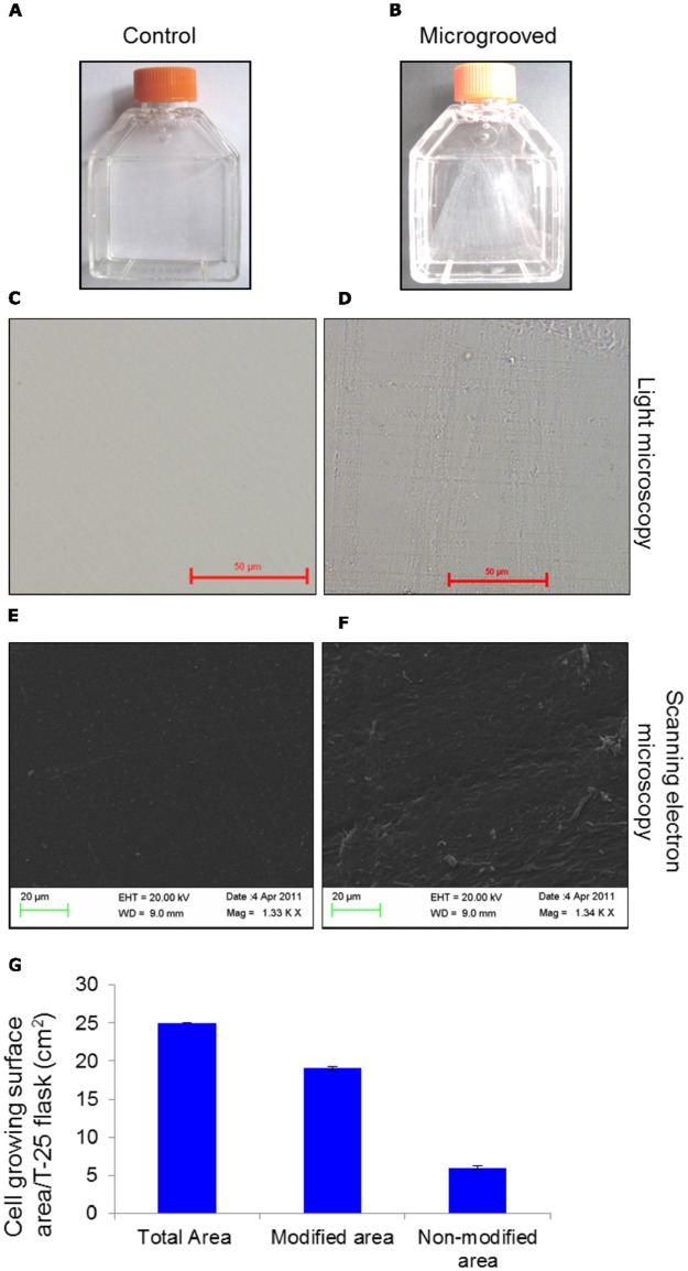
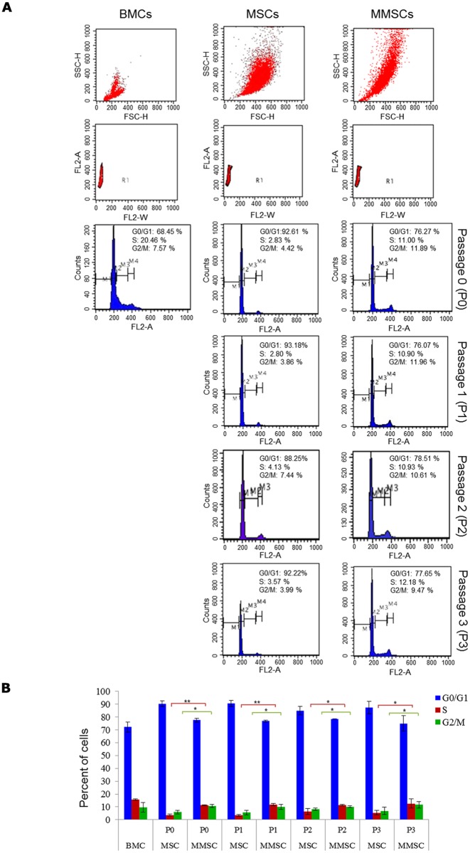
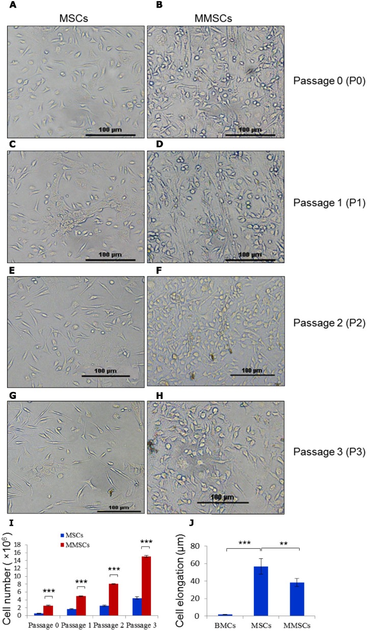
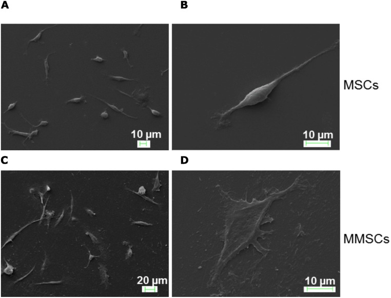
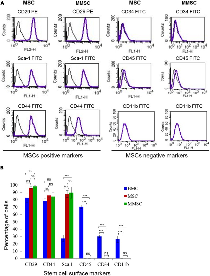
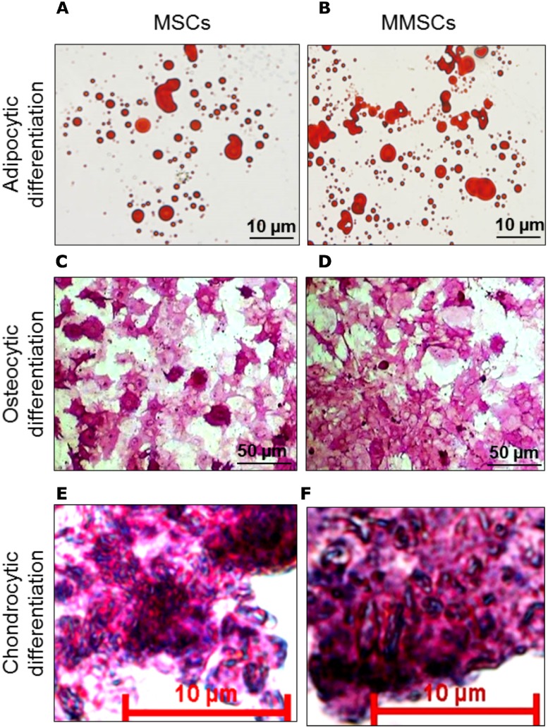
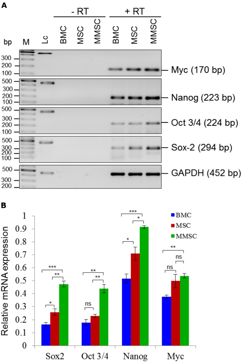
Mesenchymal stem cells' (MSCs) fate is largely determined by the various topographical features and a range of extracellular matrix (ECM) components present in their niches. Apart from maintaining structural stability, they regulate cell morphology, division, proliferation, migration and differentiation among others. Traditional MSC cultures, which are mainly based on two-dimensional smooth surfaces of culture dishes and plates, do not provide topographical cues similar to in vivo three-dimensional niches, impacting various cellular processes. Therefore, we culture the mouse bone marrow-derived MSCs on microgrooved bearing surface, partially mimicking in vivo reticulated niche, to study its effect on morphology, pluripotency factor-associated stemness, cell division and rate of proliferation. Following culture, morphological features, and MSC-specific marker gene expression, such as CD29, CD44, Sca-1 along with HSC (Haematopoietic stem cell)-specific markers like CD34, CD45, CD11b were evaluated by microscopy and immunophenotyping, respectively. HSC is another type of bone marrow stem cell population, which concertedly interacts with MSC during various functions, including haematopoiesis. In addition, mesenchymal stem cells were further analyzed for gene expression of pluripotency-associated transcription factors such as Oct3/4, Sox-2, Nanog and Myc, as well as differentiated into adipocytes, osteocytes and chondrocytes. Our results show that microgrooved surface-cultured mesenchymal stem cells (MMSCs) expressed higher levels of expected cell surface and pluripotency-associated markers and proliferated more rapidly (2-3×fold) with higher percentage of cells in S/G2-M-phase, consequently giving rise to higher cell yield compared to standard culture flask-grown cells (MSCs), taken as control. Furthermore, both MSCs and MMSCs showed considerable accumulation of intracellular lipid-droplets, higher alkaline phosphatase activity and secretion of extracellular matrix that are characteristics of adipogenesis, osteogenesis and chondrogenesis, respectively.
间充质干细胞(MSCs)的命运很大程度上取决于其微环境中存在的各种地形特征和一系列细胞外基质(ECM)成分。除了维持结构稳定性外,它们还调节细胞形态、分裂、增殖、迁移和分化等。传统的MSC培养主要基于培养皿和平板的二维光滑表面,无法提供类似于体内三维微环境的地形线索,从而影响各种细胞过程。因此,我们将小鼠骨髓来源的MSCs培养在微槽轴承表面,部分模拟体内网状微环境,以研究其对形态、多能性因子相关干性、细胞分裂和增殖速率的影响。培养后,分别通过显微镜检查和免疫表型分析评估形态特征以及MSC特异性标记基因的表达,如CD29、CD44、Sca-1以及造血干细胞(HSC)特异性标记物如CD34、CD45、CD11b。HSC是另一种骨髓干细胞群体,在包括造血在内的各种功能中与MSC协同相互作用。此外,对间充质干细胞进一步分析多能性相关转录因子如Oct3/4、Sox-2、Nanog和Myc的基因表达,并将其分化为脂肪细胞、骨细胞和软骨细胞。我们的结果表明,与作为对照的标准培养瓶培养的细胞(MSCs)相比,微槽表面培养的间充质干细胞(MMSCs)表达更高水平的预期细胞表面和多能性相关标记物,增殖更快(2 - 3倍),处于S/G2 - M期的细胞百分比更高,因此细胞产量更高。此外,MSCs和MMSCs均显示细胞内脂滴大量积累、碱性磷酸酶活性更高以及细胞外基质分泌,分别是脂肪生成、骨生成和软骨生成的特征。