• 文献检索
  • 文档翻译
  • 深度研究
  • 学术资讯
  • Suppr Zotero 插件Zotero 插件
  • 邀请有礼
  • 套餐&价格
  • 历史记录
应用&插件
Suppr Zotero 插件Zotero 插件浏览器插件Mac 客户端Windows 客户端微信小程序
定价
高级版会员购买积分包购买API积分包
服务
文献检索文档翻译深度研究API 文档MCP 服务
关于我们
关于 Suppr公司介绍联系我们用户协议隐私条款
关注我们

Suppr 超能文献

核心技术专利:CN118964589B侵权必究
粤ICP备2023148730 号-1Suppr @ 2026

文献检索

告别复杂PubMed语法,用中文像聊天一样搜索,搜遍4000万医学文献。AI智能推荐,让科研检索更轻松。

立即免费搜索

文件翻译

保留排版,准确专业,支持PDF/Word/PPT等文件格式,支持 12+语言互译。

免费翻译文档

深度研究

AI帮你快速写综述,25分钟生成高质量综述,智能提取关键信息,辅助科研写作。

立即免费体验

心肌T1映射与细胞外容积定量:技术和生物学混杂因素概述

Myocardial T1 mapping and extracellular volume quantification: an overview of technical and biological confounders.

作者信息

Piechnik Stefan K, Jerosch-Herold Michael

机构信息

Oxford Centre for Clinical Magnetic Resonance Research, Division of Cardiovascular Medicine, Radcliffe Department of Medicine, University of Oxford, John Radcliffe Hospital, Oxford, OX39DU, UK.

Brigham and Women's Hospital, and Harvard Medical School, 15 Francis Street, Boston, MA, 02115, USA.

出版信息

Int J Cardiovasc Imaging. 2018 Jan;34(1):3-14. doi: 10.1007/s10554-017-1235-7. Epub 2017 Aug 28.

DOI:10.1007/s10554-017-1235-7
PMID:28849419
原文链接:https://pmc.ncbi.nlm.nih.gov/articles/PMC5851695/
Abstract

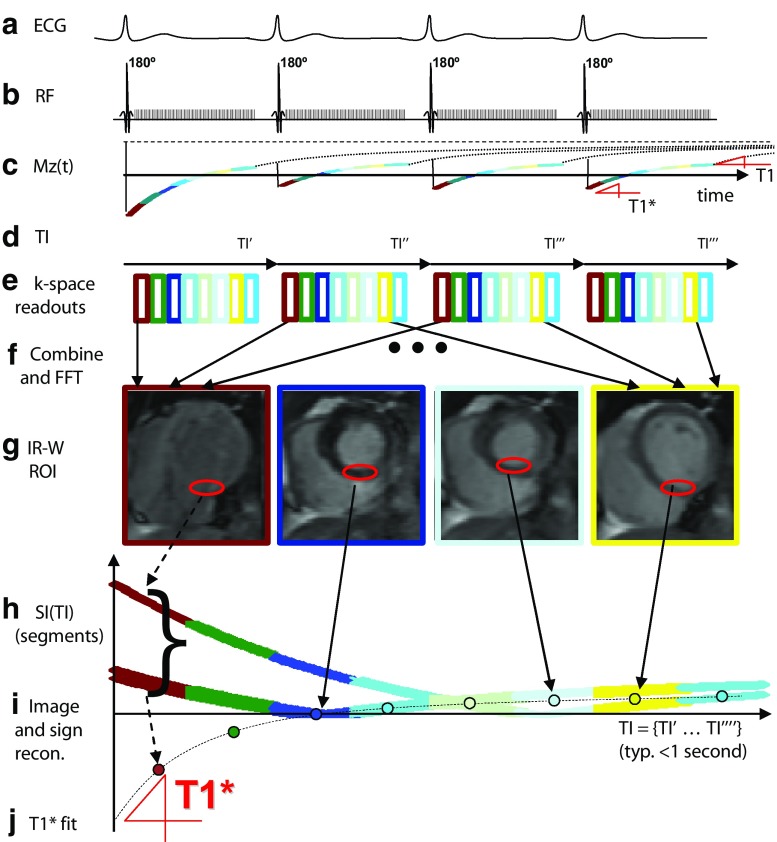
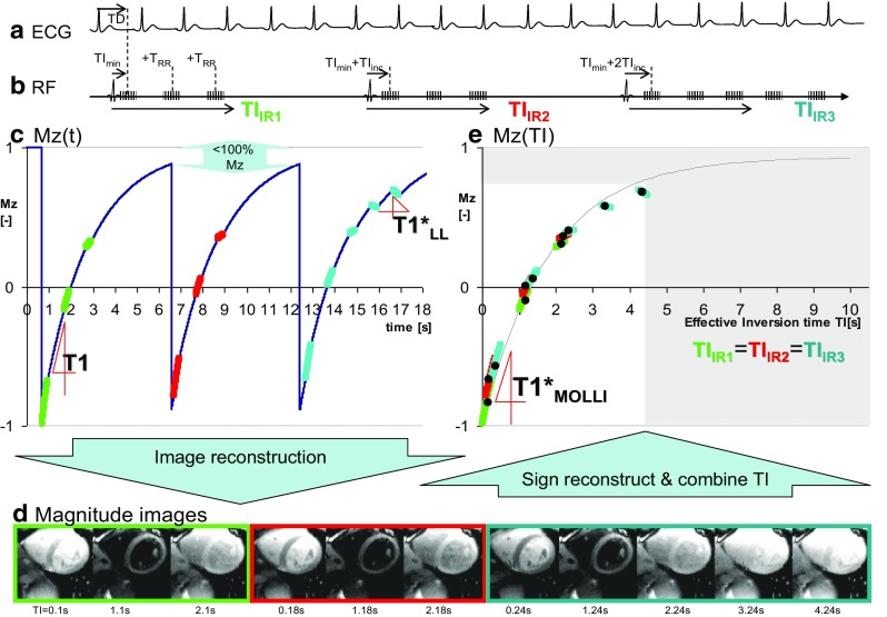
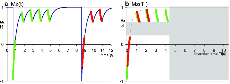
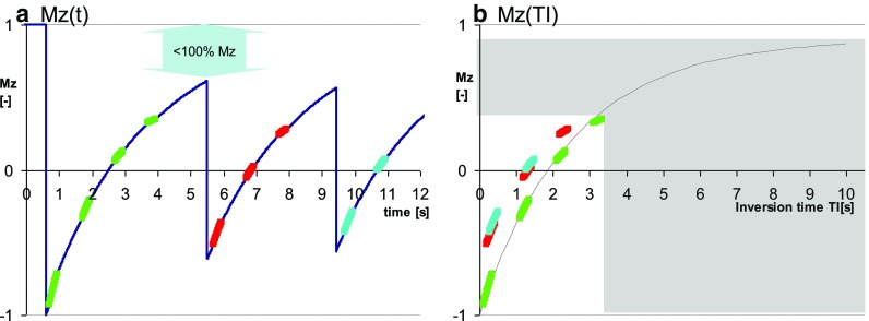
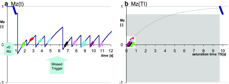
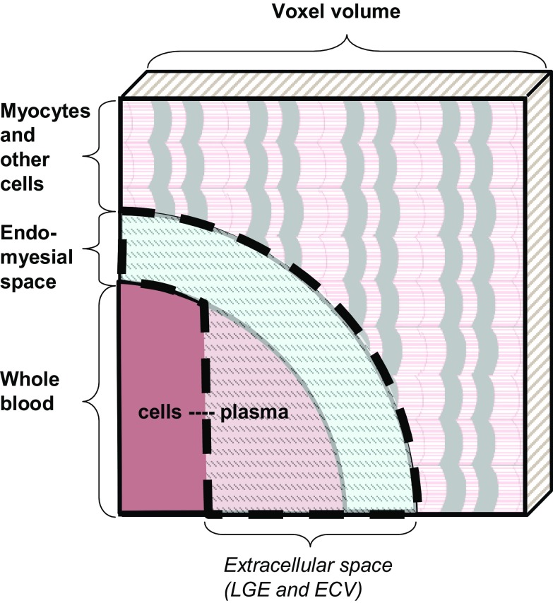
Novel tissue biomarkers based on the spin-lattice relaxation time T1, a fundamental property in the theory of magnetic resonance physics, have emerged as a new approach for myocardial tissue characterization with many validated clinical applications. This article is intended as an overview of the physical and physiological mechanisms underlying the interpretation and the accuracy of any practical measurement of T1, or derived biomarkers such as extravascular volume fraction, and also includes a discussion of potential pitfalls. Numerous caveats und knowledge gaps related to the precise interpretation of T1-based biomarkers remain, which are being addressed incrementally through ongoing research. Equally important, further careful standardization will pave the way for a wider clinical translation of these novel T1-based biomarkers of tissue remodeling, which have been well validated for their sensitivity to pathophysiological changes, though for the most part in single-center studies.

摘要

基于自旋晶格弛豫时间T1的新型组织生物标志物已成为心肌组织特征分析的一种新方法,并在许多临床应用中得到验证。T1是磁共振物理理论中的一个基本属性。本文旨在概述T1实际测量及其衍生生物标志物(如血管外容积分数)的解释和准确性背后的物理和生理机制,并讨论潜在的陷阱。与基于T1的生物标志物的精确解释相关的众多注意事项和知识空白仍然存在,目前正在通过持续研究逐步解决。同样重要的是,进一步仔细的标准化将为这些基于T1的新型组织重塑生物标志物的更广泛临床应用铺平道路。尽管在大多数单中心研究中,这些生物标志物对病理生理变化的敏感性已得到充分验证。

https://cdn.ncbi.nlm.nih.gov/pmc/blobs/0f86/5851695/5211c3791375/10554_2017_1235_Fig8_HTML.jpg
https://cdn.ncbi.nlm.nih.gov/pmc/blobs/0f86/5851695/47d87894c1c8/10554_2017_1235_Fig1_HTML.jpg
https://cdn.ncbi.nlm.nih.gov/pmc/blobs/0f86/5851695/8b5c93cf7e44/10554_2017_1235_Fig2_HTML.jpg
https://cdn.ncbi.nlm.nih.gov/pmc/blobs/0f86/5851695/e8df261a27a5/10554_2017_1235_Fig3_HTML.jpg
https://cdn.ncbi.nlm.nih.gov/pmc/blobs/0f86/5851695/16287c7df941/10554_2017_1235_Fig4_HTML.jpg
https://cdn.ncbi.nlm.nih.gov/pmc/blobs/0f86/5851695/d5ebceec99ae/10554_2017_1235_Fig5_HTML.jpg
https://cdn.ncbi.nlm.nih.gov/pmc/blobs/0f86/5851695/b23ffd94611b/10554_2017_1235_Fig6_HTML.jpg
https://cdn.ncbi.nlm.nih.gov/pmc/blobs/0f86/5851695/1a8d7e34bd00/10554_2017_1235_Fig7_HTML.jpg
https://cdn.ncbi.nlm.nih.gov/pmc/blobs/0f86/5851695/5211c3791375/10554_2017_1235_Fig8_HTML.jpg

相似文献

1
Myocardial T1 mapping and extracellular volume quantification: an overview of technical and biological confounders.心肌T1映射与细胞外容积定量:技术和生物学混杂因素概述
Int J Cardiovasc Imaging. 2018 Jan;34(1):3-14. doi: 10.1007/s10554-017-1235-7. Epub 2017 Aug 28.
2
Comprehensive validation of cardiovascular magnetic resonance techniques for the assessment of myocardial extracellular volume.全面验证心血管磁共振技术评估心肌细胞外容积。
Circ Cardiovasc Imaging. 2013 May 1;6(3):373-83. doi: 10.1161/CIRCIMAGING.112.000192. Epub 2013 Apr 3.
3
Cardiac Magnetic Resonance Fingerprinting: Technical Overview and Initial Results.心脏磁共振指纹技术:技术概述与初步结果。
JACC Cardiovasc Imaging. 2018 Dec;11(12):1837-1853. doi: 10.1016/j.jcmg.2018.08.028.
4
T1 Mapping: Basic Techniques and Clinical Applications.T1 映射:基本技术与临床应用。
JACC Cardiovasc Imaging. 2016 Jan;9(1):67-81. doi: 10.1016/j.jcmg.2015.11.005.
5
Advances in parametric mapping with CMR imaging.CMR 成像的参数图研究进展。
JACC Cardiovasc Imaging. 2013 Jul;6(7):806-22. doi: 10.1016/j.jcmg.2013.05.005.
6
Automatic Measurement of the Myocardial Interstitium: Synthetic Extracellular Volume Quantification Without Hematocrit Sampling.心肌间质的自动测量:无需红细胞压积采样的合成细胞外体积定量。
JACC Cardiovasc Imaging. 2016 Jan;9(1):54-63. doi: 10.1016/j.jcmg.2015.11.008.
7
Association between extracellular matrix expansion quantified by cardiovascular magnetic resonance and short-term mortality.心血管磁共振量化的细胞外基质扩张与短期死亡率的关系。
Circulation. 2012 Sep 4;126(10):1206-16. doi: 10.1161/CIRCULATIONAHA.111.089409. Epub 2012 Jul 31.
8
Myocardial T1 and ECV Measurement: Underlying Concepts and Technical Considerations.心肌 T1 和 ECV 测量:基本概念和技术考虑因素。
JACC Cardiovasc Imaging. 2019 Nov;12(11 Pt 2):2332-2344. doi: 10.1016/j.jcmg.2019.06.031. Epub 2019 Sep 18.
9
T1 mapping of the myocardium: intra-individual assessment of post-contrast T1 time evolution and extracellular volume fraction at 3T for Gd-DTPA and Gd-BOPTA.心肌 T1 映射:3T 下钆-DTPA 和钆-BOPTA 对比后 T1 时间演变和细胞外容积分数的个体内评估。
J Cardiovasc Magn Reson. 2012 Apr 28;14(1):26. doi: 10.1186/1532-429X-14-26.
10
Importance of standardizing timing of hematocrit measurement when using cardiovascular magnetic resonance to calculate myocardial extracellular volume (ECV) based on pre- and post-contrast T1 mapping.使用心血管磁共振基于钆对比前后 T1 映射计算心肌细胞外容积(ECV)时,标准化红细胞比容测量时间的重要性。
J Cardiovasc Magn Reson. 2018 Jun 28;20(1):46. doi: 10.1186/s12968-018-0464-9.

引用本文的文献

1
Generalizable, sequence-invariant deep learning image reconstruction for subspace-constrained quantitative MRI.用于子空间约束定量磁共振成像的可推广、序列不变深度学习图像重建
Magn Reson Med. 2025 Jul;94(1):89-104. doi: 10.1002/mrm.30433. Epub 2025 Jan 20.
2
Cardiac magnetic resonance imaging for discrimination of hypertensive heart disease and hypertrophic cardiomyopathy: a systematic review and meta-analysis.心脏磁共振成像用于鉴别高血压性心脏病和肥厚型心肌病:一项系统评价和荟萃分析。
Front Cardiovasc Med. 2024 Aug 2;11:1421013. doi: 10.3389/fcvm.2024.1421013. eCollection 2024.
3
Assessing the Volume of the Head of the Mandibular Condyle Using 3T-MRI-A Preliminary Trial.

本文引用的文献

1
Synthetic hematocrit derived from the longitudinal relaxation of blood can lead to clinically significant errors in measurement of extracellular volume fraction in pediatric and young adult patients.源自血液纵向弛豫的合成血细胞比容可能导致儿科和年轻成年患者细胞外体积分数测量出现具有临床意义的误差。
J Cardiovasc Magn Reson. 2017 Aug 2;19(1):58. doi: 10.1186/s12968-017-0377-z.
2
Splenic T1-mapping: a novel quantitative method for assessing adenosine stress adequacy for cardiovascular magnetic resonance.脾脏T1映射:一种评估心血管磁共振腺苷负荷充足性的新型定量方法。
J Cardiovasc Magn Reson. 2017 Jan 13;19(1):1. doi: 10.1186/s12968-016-0318-2.
3
使用3T磁共振成像评估下颌髁突头部体积——一项初步试验
Dent J (Basel). 2024 Jul 16;12(7):220. doi: 10.3390/dj12070220.
4
Quantitating myocardial fibrosis using extracellular extravascular volume determined from computed tomography myocardial perfusion imaging.利用计算机断层心肌灌注成像确定的细胞外细胞外体积定量心肌纤维化。
BMC Med Imaging. 2024 Feb 12;24(1):40. doi: 10.1186/s12880-024-01226-3.
5
Early myocardial oedema can predict subsequent cardiomyopathy in high-dose anthracycline therapy.早期心肌水肿可预测大剂量蒽环类药物治疗后继发心肌病。
ESC Heart Fail. 2023 Feb;10(1):616-627. doi: 10.1002/ehf2.14232. Epub 2022 Nov 20.
6
Molecular Approaches and Echocardiographic Deformation Imaging in Detecting Myocardial Fibrosis.分子方法和超声心动图应变成像在心肌纤维化检测中的应用。
Int J Mol Sci. 2022 Sep 19;23(18):10944. doi: 10.3390/ijms231810944.
7
MOCOnet: Robust Motion Correction of Cardiovascular Magnetic Resonance T1 Mapping Using Convolutional Neural Networks.MOCOnet:使用卷积神经网络对心血管磁共振T1映射进行稳健的运动校正
Front Cardiovasc Med. 2021 Nov 23;8:768245. doi: 10.3389/fcvm.2021.768245. eCollection 2021.
8
Deep learning with attention supervision for automated motion artefact detection in quality control of cardiac T1-mapping.用于心脏T1映射质量控制中自动运动伪影检测的带注意力监督的深度学习
Artif Intell Med. 2020 Nov;110:101955. doi: 10.1016/j.artmed.2020.101955. Epub 2020 Sep 7.
9
Influence of hydration status on cardiovascular magnetic resonance myocardial T1 and T2 relaxation time assessment: an intraindividual study in healthy subjects.探讨水合状态对心血管磁共振心肌 T1 和 T2 弛豫时间评估的影响:健康受试者的个体内研究。
J Cardiovasc Magn Reson. 2020 Sep 7;22(1):63. doi: 10.1186/s12968-020-00661-9.
10
Cardiovascular Magnetic Resonance for the Differentiation of Left Ventricular Hypertrophy.心血管磁共振在左心室肥厚鉴别诊断中的应用。
Curr Heart Fail Rep. 2020 Oct;17(5):192-204. doi: 10.1007/s11897-020-00481-z.
Myocardial T-mapping at 3T using saturation-recovery: reference values, precision and comparison with MOLLI.
使用饱和恢复法在3T下进行心肌T值映射:参考值、精密度及与MOLLI的比较
J Cardiovasc Magn Reson. 2016 Nov 18;18(1):84. doi: 10.1186/s12968-016-0302-x.
4
Comparison of different cardiovascular magnetic resonance sequences for native myocardial T1 mapping at 3T.3T下用于天然心肌T1映射的不同心血管磁共振序列的比较
J Cardiovasc Magn Reson. 2016 Oct 4;18(1):65. doi: 10.1186/s12968-016-0286-6.
5
Robust free-breathing SASHA T1 mapping with high-contrast image registration.基于高对比度图像配准的稳健自由呼吸SASHA T1映射
J Cardiovasc Magn Reson. 2016 Aug 17;18(1):47. doi: 10.1186/s12968-016-0267-9.
6
Caffeine intake inverts the effect of adenosine on myocardial perfusion during stress as measured by T1 mapping.通过T1映射测量发现,在应激状态下,咖啡因摄入会逆转腺苷对心肌灌注的影响。
Int J Cardiovasc Imaging. 2016 Oct;32(10):1545-53. doi: 10.1007/s10554-016-0949-2. Epub 2016 Jul 29.
7
Automatic Measurement of the Myocardial Interstitium: Synthetic Extracellular Volume Quantification Without Hematocrit Sampling.心肌间质的自动测量:无需红细胞压积采样的合成细胞外体积定量。
JACC Cardiovasc Imaging. 2016 Jan;9(1):54-63. doi: 10.1016/j.jcmg.2015.11.008.
8
The Behavior of Water in Collagen and Hydroxyapatite Sites of Cortical Bone: Fracture, Mechanical Wear, and Load Bearing Studies.皮质骨中胶原蛋白和羟基磷灰石部位的水行为:骨折、机械磨损和承重研究。
J Phys Chem C Nanomater Interfaces. 2015 Sep 17;119(37):21528-21537. doi: 10.1021/acs.jpcc.5b06285. Epub 2015 Aug 28.
9
Saturation pulse design for quantitative myocardial T1 mapping.用于定量心肌T1映射的饱和脉冲设计
J Cardiovasc Magn Reson. 2015 Oct 1;17:84. doi: 10.1186/s12968-015-0187-0.
10
Dose correction for post-contrast T1 mapping of the heart: the MESA study.心脏对比剂增强T1映射的剂量校正:多民族动脉粥样硬化研究(MESA研究)
Int J Cardiovasc Imaging. 2016 Feb;32(2):271-279. doi: 10.1007/s10554-015-0754-3. Epub 2015 Sep 11.