Colman Michael A, Pinali Christian, Trafford Andrew W, Zhang Henggui, Kitmitto Ashraf
School of Biomedical Sciences, Faculty of Biological Sciences, University of Leeds, Leeds, United Kingdom.
School of Physics and Astronomy, Faculty of Engineering and Physical Sciences, University of Manchester, Manchester, United Kingdom.
PLoS Comput Biol. 2017 Aug 31;13(8):e1005714. doi: 10.1371/journal.pcbi.1005714. eCollection 2017 Aug.
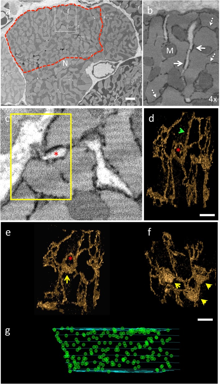
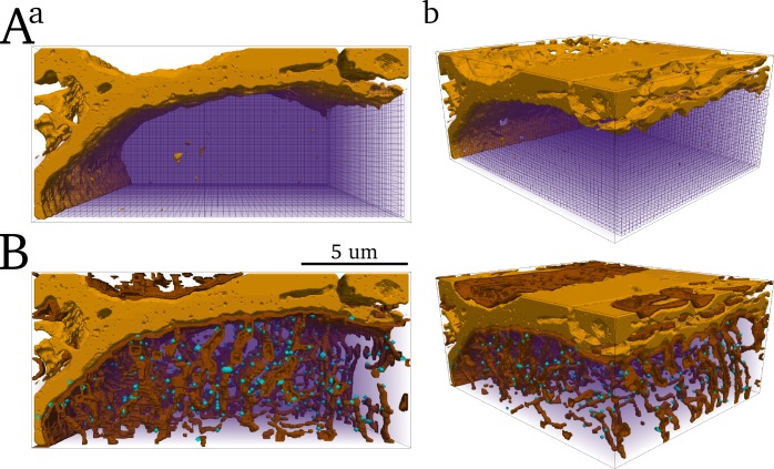
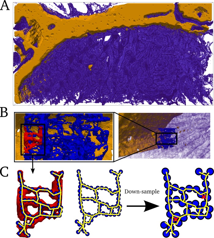
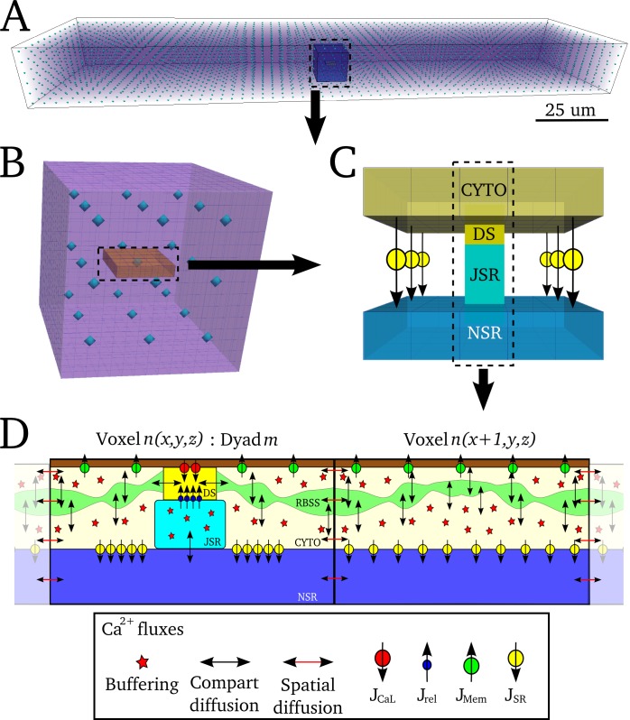
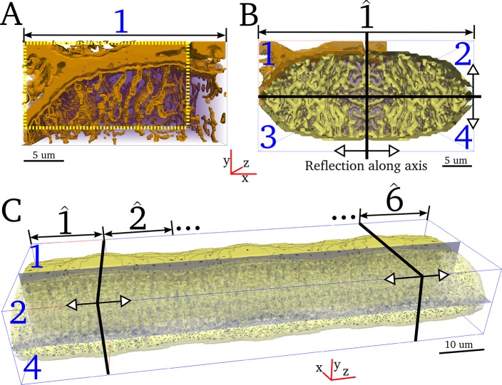
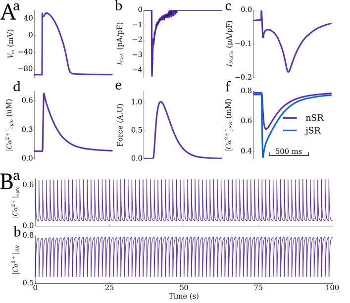
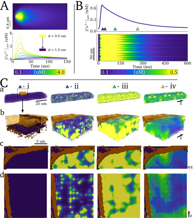
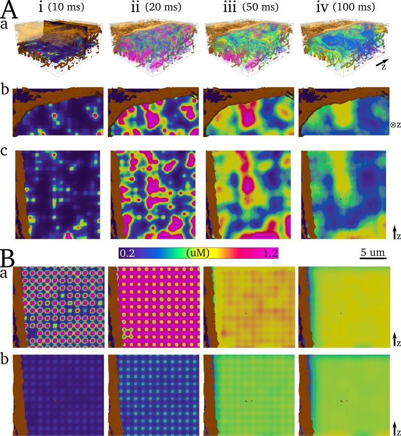
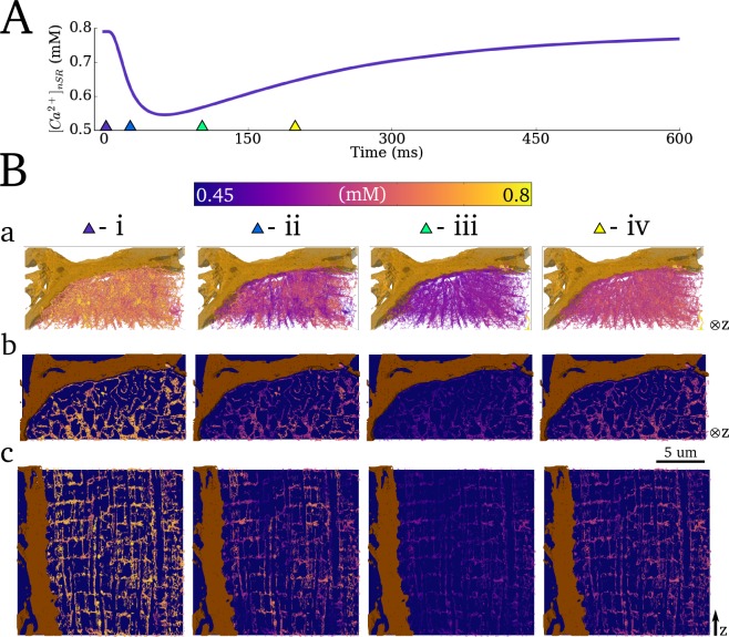
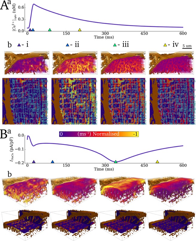
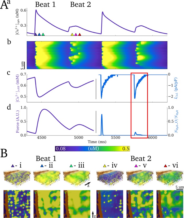
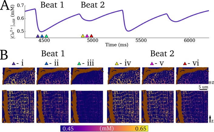
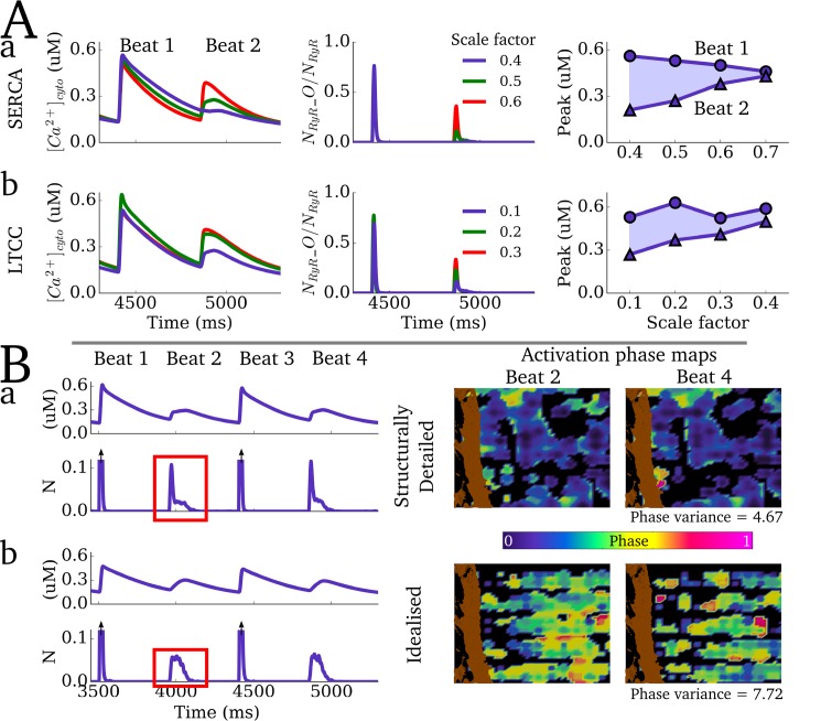
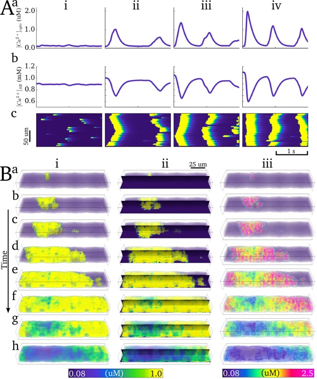
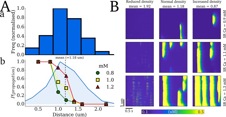
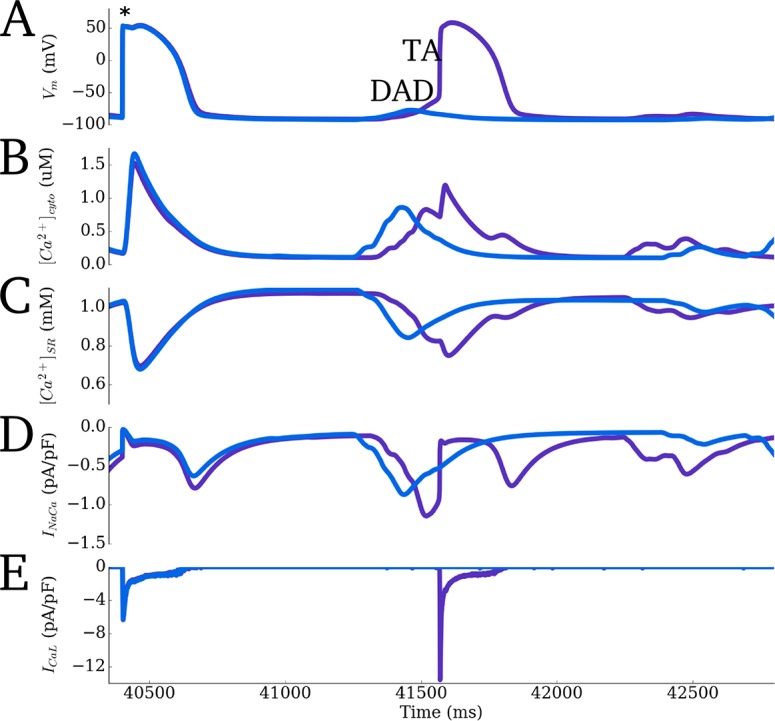
Intracellular calcium cycling is a vital component of cardiac excitation-contraction coupling. The key structures responsible for controlling calcium dynamics are the cell membrane (comprising the surface sarcolemma and transverse-tubules), the intracellular calcium store (the sarcoplasmic reticulum), and the co-localisation of these two structures to form dyads within which calcium-induced-calcium-release occurs. The organisation of these structures tightly controls intracellular calcium dynamics. In this study, we present a computational model of intracellular calcium cycling in three-dimensions (3-D), which incorporates high resolution reconstructions of these key regulatory structures, attained through imaging of tissue taken from the sheep left ventricle using serial block face scanning electron microscopy. An approach was developed to model the sarcoplasmic reticulum structure at the whole-cell scale, by reducing its full 3-D structure to a 3-D network of one-dimensional strands. The model reproduces intracellular calcium dynamics during control pacing and reveals the high-resolution 3-D spatial structure of calcium gradients and intracellular fluxes in both the cytoplasm and sarcoplasmic reticulum. We also demonstrated the capability of the model to reproduce potentially pro-arrhythmic dynamics under perturbed conditions, pertaining to calcium-transient alternans and spontaneous release events. Comparison with idealised cell models emphasised the importance of structure in determining calcium gradients and controlling the spatial dynamics associated with calcium-transient alternans, wherein the probabilistic nature of dyad activation and recruitment was constrained. The model was further used to highlight the criticality in calcium spark propagation in relation to inter-dyad distances. The model presented provides a powerful tool for future investigation of structure-function relationships underlying physiological and pathophysiological intracellular calcium handling phenomena at the whole-cell. The approach allows for the first time direct integration of high-resolution images of 3-D intracellular structures with models of calcium cycling, presenting the possibility to directly assess the functional impact of structural remodelling at the cellular scale.
细胞内钙循环是心脏兴奋-收缩偶联的重要组成部分。负责控制钙动力学的关键结构是细胞膜(包括表面肌膜和横管)、细胞内钙库(肌浆网),以及这两种结构的共定位形成二联体,在二联体内发生钙诱导钙释放。这些结构的组织紧密控制着细胞内钙动力学。在本研究中,我们提出了一个三维(3-D)细胞内钙循环计算模型,该模型纳入了这些关键调节结构的高分辨率重建,这些重建是通过使用连续块面扫描电子显微镜对取自绵羊左心室的组织进行成像获得的。开发了一种方法,通过将肌浆网的完整三维结构简化为一维链的三维网络,在全细胞尺度上对其结构进行建模。该模型再现了对照起搏期间的细胞内钙动力学,并揭示了细胞质和肌浆网中钙梯度和细胞内通量的高分辨率三维空间结构。我们还证明了该模型在受扰条件下再现潜在致心律失常动力学的能力,这些条件与钙瞬变交替和自发释放事件有关。与理想化细胞模型的比较强调了结构在确定钙梯度和控制与钙瞬变交替相关的空间动力学方面的重要性,其中二联体激活和募集的概率性质受到限制。该模型还被用于强调钙火花传播相对于二联体间距离的关键性。所提出的模型为未来研究全细胞生理和病理生理细胞内钙处理现象背后的结构-功能关系提供了一个强大的工具。该方法首次允许将三维细胞内结构的高分辨率图像与钙循环模型直接整合,从而有可能在细胞尺度上直接评估结构重塑的功能影响。