Graumans Wouter, Tadesse Fitsum G, Andolina Chiara, van Gemert Geert-Jan, Teelen Karina, Lanke Kjerstin, Gadisa Endalamaw, Yewhalaw Delenasaw, van de Vegte-Bolmer Marga, Siebelink-Stoter Rianne, Reuling Isaïe, Sauerwein Robert, Bousema Teun
Department of Medical Microbiology, Radboud University Nijmegen Medical Centre, Nijmegen, The Netherlands.
Armauer Hansen Research Institute (AHRI), Addis Ababa, Ethiopia.
Malar J. 2017 Sep 6;16(1):356. doi: 10.1186/s12936-017-2011-9.
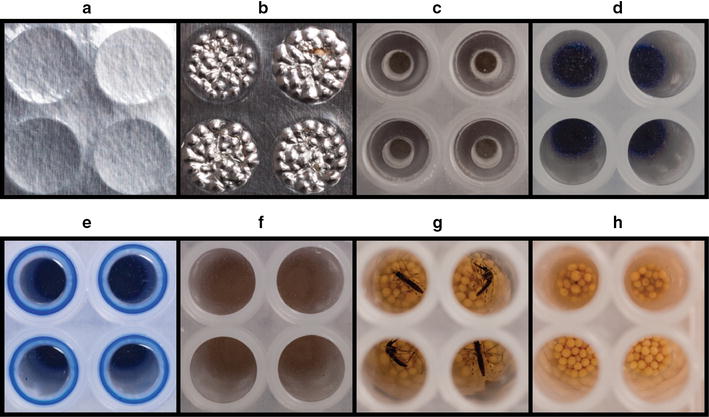
The malaria infection status of mosquitoes is commonly determined by microscopic detection of oocysts on the dissected mosquito midgut. This method is labour-intensive, does not allow processing of large numbers of mosquitoes and can be challenging in terms of objective classification of oocysts. Here, a semi-high-throughput bead-beating ELISA method is proposed for detection of the circumsporozoite protein (CSP) followed by confirmation by quantitative PCR (qPCR).
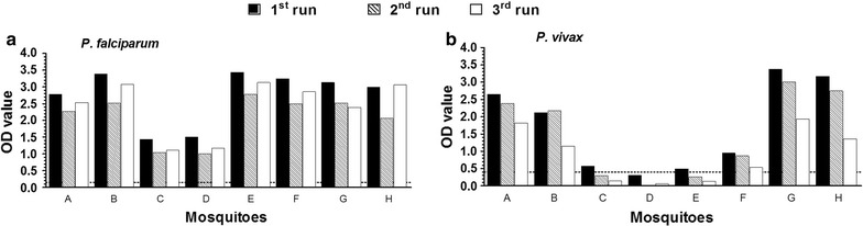
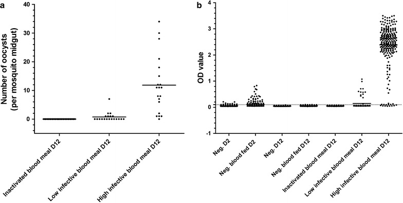
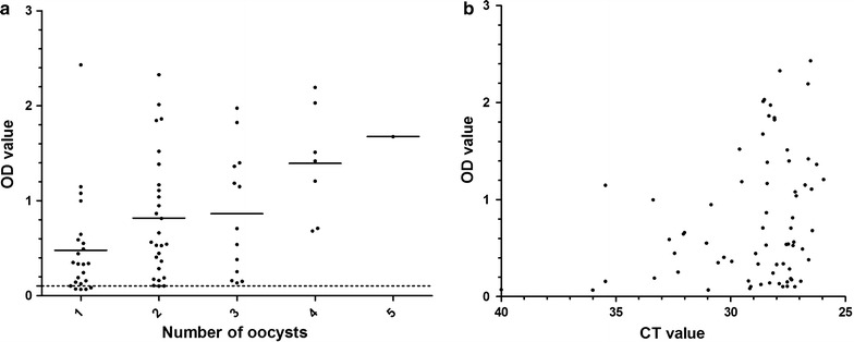
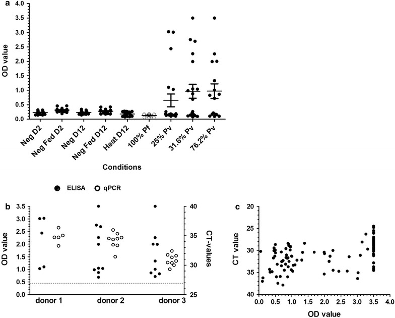
Cultured Plasmodium falciparum gametocytes were offered to Anopheles stephensi mosquitoes and examined by microscopy. After bead-beating, mosquito homogenate was examined by CSP-ELISA and 18S qPCR. As negative controls, mosquitoes that were offered a heat-inactivated gametocyte blood meal were used. The CSP-ELISA/qPCR methodology was applied to high and low-intensity infections of cultured P. falciparum gametocytes. A similar methodology optimized for P. vivax was used on mosquitoes that were offered blood from Ethiopian donors who were naturally infected with P. vivax.
There was considerable variation in CSP-ELISA signal and qPCR values in mosquitoes with low oocyst intensities. There was a strong agreement mosquito positivity by CSP-ELISA and by qPCR in mosquitoes that fed on cultured P. falciparum material (agreement 96.9%; kappa = 0.97) and naturally infected P. vivax parasite carriers [agreement 92.4% (kappa = 0.83)].
The proposed bead-beating CSP-ELISA/qPCR methodology considerably increases throughput for the detection of mosquito infection. qPCR remains necessary to confirm infections in mosquitoes with low CSP-ELISA signal. This methodology may prove particularly useful for studies where very low mosquito infection prevalence is expected and study sites where experience with oocyst detection is limited.
蚊子的疟疾感染状况通常通过显微镜检测解剖后的蚊子中肠上的卵囊来确定。这种方法劳动强度大,无法处理大量蚊子,并且在卵囊的客观分类方面可能具有挑战性。在此,提出了一种半高通量的珠磨酶联免疫吸附测定(ELISA)方法来检测环子孢子蛋白(CSP),随后通过定量聚合酶链反应(qPCR)进行确认。
将培养的恶性疟原虫配子体提供给斯氏按蚊,并通过显微镜检查。珠磨后,通过CSP-ELISA和18S qPCR检测蚊子匀浆。作为阴性对照,使用喂食热灭活配子体血餐的蚊子。CSP-ELISA/qPCR方法应用于培养的恶性疟原虫配子体的高强度和低强度感染。一种针对间日疟原虫优化的类似方法用于喂食来自自然感染间日疟原虫的埃塞俄比亚献血者血液的蚊子。
卵囊强度低的蚊子中,CSP-ELISA信号和qPCR值存在相当大的差异。在喂食培养的恶性疟原虫材料的蚊子中,CSP-ELISA和qPCR检测蚊子阳性的一致性很强(一致性96.9%;kappa = 0.97),在自然感染间日疟原虫的寄生虫携带者中也是如此[一致性92.4%(kappa = 0.83)]。
所提出的珠磨CSP-ELISA/qPCR方法大大提高了蚊子感染检测的通量。对于CSP-ELISA信号低的蚊子感染,仍需要qPCR来确认。这种方法可能对预期蚊子感染率非常低的研究以及卵囊检测经验有限的研究地点特别有用。