• 文献检索
  • 文档翻译
  • 深度研究
  • 学术资讯
  • Suppr Zotero 插件Zotero 插件
  • 邀请有礼
  • 套餐&价格
  • 历史记录
应用&插件
Suppr Zotero 插件Zotero 插件浏览器插件Mac 客户端Windows 客户端微信小程序
定价
高级版会员购买积分包购买API积分包
服务
文献检索文档翻译深度研究API 文档MCP 服务
关于我们
关于 Suppr公司介绍联系我们用户协议隐私条款
关注我们

Suppr 超能文献

核心技术专利:CN118964589B侵权必究
粤ICP备2023148730 号-1Suppr @ 2026

文献检索

告别复杂PubMed语法,用中文像聊天一样搜索,搜遍4000万医学文献。AI智能推荐,让科研检索更轻松。

立即免费搜索

文件翻译

保留排版,准确专业,支持PDF/Word/PPT等文件格式,支持 12+语言互译。

免费翻译文档

深度研究

AI帮你快速写综述,25分钟生成高质量综述,智能提取关键信息,辅助科研写作。

立即免费体验

人体血液中类似多形细菌的结构代表无生命的膜泡和蛋白颗粒。

Pleomorphic bacteria-like structures in human blood represent non-living membrane vesicles and protein particles.

机构信息

Center for Molecular and Clinical Immunology, Chang Gung University, Taoyuan, Taiwan.

Laboratory of Nanomaterials, Chang Gung University, Taoyuan, Taiwan.

出版信息

Sci Rep. 2017 Sep 6;7(1):10650. doi: 10.1038/s41598-017-10479-8.

DOI:10.1038/s41598-017-10479-8
PMID:28878382
原文链接:https://pmc.ncbi.nlm.nih.gov/articles/PMC5587737/
Abstract

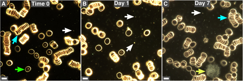
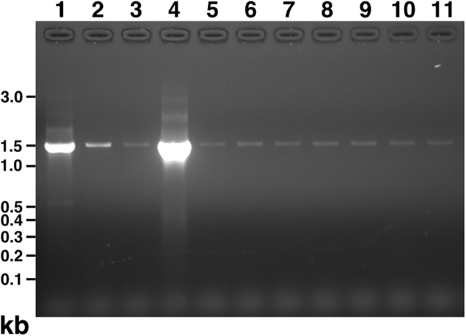
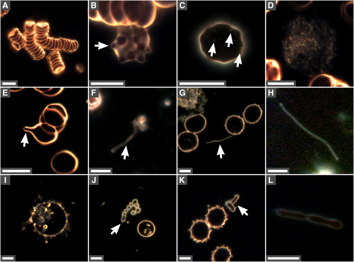
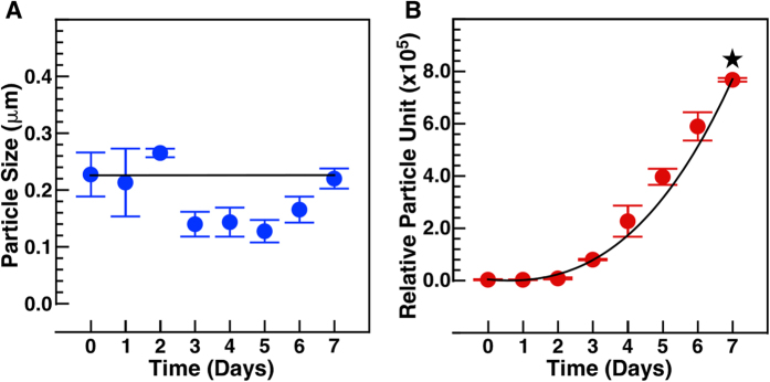
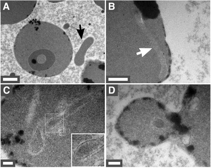
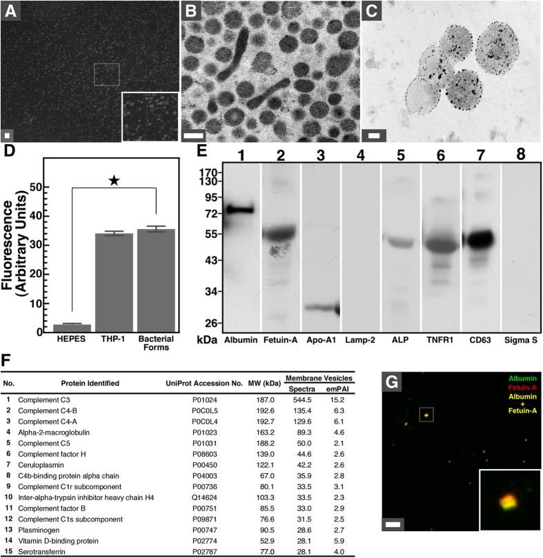
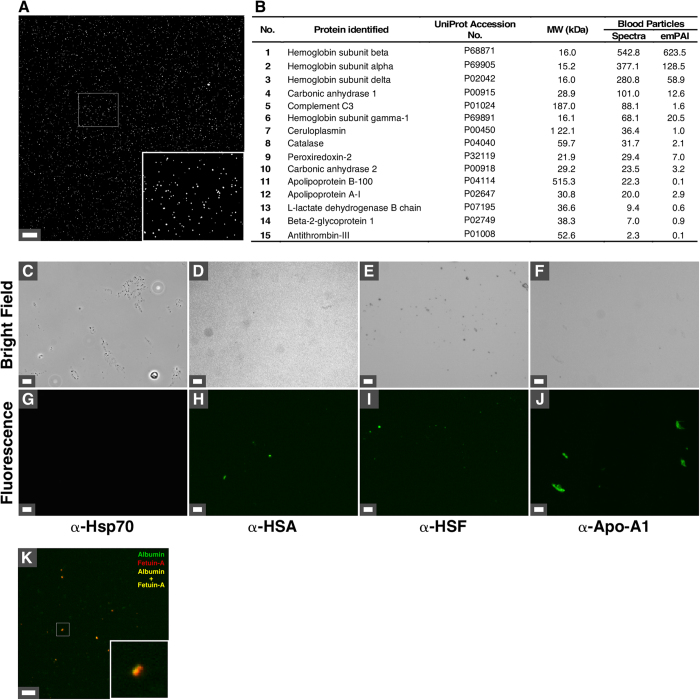
Although human blood is believed to be a sterile environment, recent studies suggest that pleomorphic bacteria exist in the blood of healthy humans. These studies have led to the development of "live-blood analysis," a technique used by alternative medicine practitioners to diagnose various human conditions, including allergies, cancer, cardiovascular disease and septicemia. We show here that bacteria-like vesicles and refringent particles form in healthy human blood observed under dark-field microscopy. These structures gradually increase in number during incubation and show morphologies reminiscent of cells undergoing division. Based on lipid analysis and Western blotting, we show that the bacteria-like entities consist of membrane vesicles containing serum and exosome proteins, including albumin, fetuin-A, apolipoprotein-A1, alkaline phosphatase, TNFR1 and CD63. In contrast, the refringent particles represent protein aggregates that contain several blood proteins. 16S rDNA PCR analysis reveals the presence of bacterial DNA in incubated blood samples but also in negative controls, indicating that the amplified sequences represent contaminants. These results suggest that the bacteria-like vesicles and refringent particles observed in human blood represent non-living membrane vesicles and protein aggregates derived from blood. The phenomena observed during live-blood analysis are therefore consistent with time-dependent decay of cells and body fluids during incubation ex vivo.

摘要

尽管人们认为血液是无菌的环境,但最近的研究表明,多形细菌存在于健康人体的血液中。这些研究导致了“活体血液分析”的发展,这是一种替代医学从业者用来诊断各种人类疾病的技术,包括过敏、癌症、心血管疾病和败血症。我们在这里展示,在暗场显微镜下观察到健康人血液中形成了类似细菌的囊泡和折射颗粒。这些结构在孵育过程中数量逐渐增加,并表现出类似于正在分裂的细胞的形态。基于脂质分析和 Western blot,我们表明类似细菌的实体由含有血清和外泌体蛋白的膜囊泡组成,包括白蛋白、胎球蛋白-A、载脂蛋白-A1、碱性磷酸酶、TNFR1 和 CD63。相比之下,折射颗粒代表含有几种血液蛋白的蛋白质聚集体。16S rDNA PCR 分析显示孵育血液样本中存在细菌 DNA,但阴性对照中也存在,表明扩增序列代表污染物。这些结果表明,在人血液中观察到的类似细菌的囊泡和折射颗粒代表源自血液的非生活膜囊泡和蛋白质聚集体。因此,活体血液分析中观察到的现象与体外孵育过程中细胞和体液的时间依赖性衰减是一致的。

https://cdn.ncbi.nlm.nih.gov/pmc/blobs/2a7b/5587737/fc74c5ada54c/41598_2017_10479_Fig7_HTML.jpg
https://cdn.ncbi.nlm.nih.gov/pmc/blobs/2a7b/5587737/b989ad08db6b/41598_2017_10479_Fig1_HTML.jpg
https://cdn.ncbi.nlm.nih.gov/pmc/blobs/2a7b/5587737/7a7295eba316/41598_2017_10479_Fig2_HTML.jpg
https://cdn.ncbi.nlm.nih.gov/pmc/blobs/2a7b/5587737/4fb182250b50/41598_2017_10479_Fig3_HTML.jpg
https://cdn.ncbi.nlm.nih.gov/pmc/blobs/2a7b/5587737/cba33ff4534b/41598_2017_10479_Fig4_HTML.jpg
https://cdn.ncbi.nlm.nih.gov/pmc/blobs/2a7b/5587737/93d4901eeeaa/41598_2017_10479_Fig5_HTML.jpg
https://cdn.ncbi.nlm.nih.gov/pmc/blobs/2a7b/5587737/b25055072642/41598_2017_10479_Fig6_HTML.jpg
https://cdn.ncbi.nlm.nih.gov/pmc/blobs/2a7b/5587737/fc74c5ada54c/41598_2017_10479_Fig7_HTML.jpg

相似文献

1
Pleomorphic bacteria-like structures in human blood represent non-living membrane vesicles and protein particles.人体血液中类似多形细菌的结构代表无生命的膜泡和蛋白颗粒。
Sci Rep. 2017 Sep 6;7(1):10650. doi: 10.1038/s41598-017-10479-8.
2
Proteomic characterization of macro-, micro- and nano-extracellular vesicles derived from the same first trimester placenta: relevance for feto-maternal communication.从同一妊娠早期胎盘来源的大、小、纳米细胞外囊泡中进行蛋白质组学表征:与胎母通讯的相关性。
Hum Reprod. 2016 Apr;31(4):687-99. doi: 10.1093/humrep/dew004. Epub 2016 Feb 1.
3
Membrane vesicles nucleate mineralo-organic nanoparticles and induce carbonate apatite precipitation in human body fluids.膜泡在人体体液中诱导矿化有机纳米颗粒的形成和碳酸磷灰石的沉淀。
J Biol Chem. 2013 Oct 18;288(42):30571-30584. doi: 10.1074/jbc.M113.492157. Epub 2013 Aug 29.
4
Production of extracellular vesicles with light-induced proton pump activity by proteorhodopsin-containing marine bacteria.利用含菌视紫红质的海洋细菌产生具有光诱导质子泵活性的细胞外囊泡。
Microbiologyopen. 2019 Aug;8(8):e00808. doi: 10.1002/mbo3.808. Epub 2019 Feb 22.
5
Characterization of Nanohybridosomes from Lipids and Spruce Homogenate Containing Extracellular Vesicles.从含细胞外囊泡的脂类和云杉匀浆中提取纳米杂合囊泡的特性分析。
Int J Nanomedicine. 2024 Feb 22;19:1709-1721. doi: 10.2147/IJN.S432836. eCollection 2024.
6
A comparison of methods for the isolation and separation of extracellular vesicles from protein and lipid particles in human serum.比较从人血清中蛋白质和脂质颗粒中分离和分离细胞外囊泡的方法。
Sci Rep. 2020 Jan 23;10(1):1039. doi: 10.1038/s41598-020-57497-7.
7
Streptococcal Extracellular Membrane Vesicles Are Rapidly Internalized by Immune Cells and Alter Their Cytokine Release.链球菌细胞外膜囊泡被免疫细胞快速内吞,并改变其细胞因子释放。
Front Immunol. 2020 Feb 14;11:80. doi: 10.3389/fimmu.2020.00080. eCollection 2020.
8
Detailed analysis of the plasma extracellular vesicle proteome after separation from lipoproteins.详细分析脂蛋白分离后的血浆细胞外囊泡蛋白质组。
Cell Mol Life Sci. 2018 Aug;75(15):2873-2886. doi: 10.1007/s00018-018-2773-4. Epub 2018 Feb 13.
9
Cryo-electron microscopy of extracellular vesicles from cerebrospinal fluid.脑脊液细胞外囊泡的冷冻电子显微镜观察
PLoS One. 2020 Jan 30;15(1):e0227949. doi: 10.1371/journal.pone.0227949. eCollection 2020.
10
Methods to Analyze EVs.分析细胞外囊泡的方法。
Methods Mol Biol. 2017;1545:1-20. doi: 10.1007/978-1-4939-6728-5_1.

引用本文的文献

1
Blood microbiome signatures in systemic diseases: current insights, methodological pitfalls, and future horizons.全身性疾病中的血液微生物组特征:当前见解、方法学陷阱及未来展望
Front Cell Infect Microbiol. 2025 Jul 28;15:1616029. doi: 10.3389/fcimb.2025.1616029. eCollection 2025.
2
Choice of blood collection methods influences extracellular vesicles counts and miRNA profiling.采血方法的选择会影响细胞外囊泡计数和微小RNA谱分析。
J Extracell Biol. 2024 Oct 22;3(10):e70008. doi: 10.1002/jex2.70008. eCollection 2024 Oct.
3
Blood Microbiota and Its Products: Mechanisms of Interference with Host Cells and Clinical Outcomes.

本文引用的文献

1
Translocation of mineralo-organic nanoparticles from blood to urine: a new mechanism for the formation of kidney stones?矿物质-有机纳米颗粒从血液到尿液的转移:肾结石形成的新机制?
Nanomedicine (Lond). 2016 Sep;11(18):2399-404. doi: 10.2217/nnm-2016-0246. Epub 2016 Aug 8.
2
Fatty acids and small organic compounds bind to mineralo-organic nanoparticles derived from human body fluids as revealed by metabolomic analysis.代谢组学分析显示,脂肪酸和小有机化合物与源自人体体液的矿质有机纳米颗粒结合。
Nanoscale. 2016 Mar 14;8(10):5537-45. doi: 10.1039/c5nr08116e.
3
Detection and characterization of mineralo-organic nanoparticles in human kidneys.
血液微生物群及其产物:干扰宿主细胞的机制与临床结局
Hematol Rep. 2024 Jul 6;16(3):440-453. doi: 10.3390/hematolrep16030043.
4
The Blood Microbiome and Health: Current Evidence, Controversies, and Challenges.血液微生物组与健康:当前的证据、争议和挑战。
Int J Mol Sci. 2023 Mar 15;24(6):5633. doi: 10.3390/ijms24065633.
5
Morphology of blood microbiota in healthy individuals assessed by light and electron microscopy.采用光镜和电子显微镜评估健康个体血液微生物群的形态。
Front Cell Infect Microbiol. 2023 Jan 18;12:1091341. doi: 10.3389/fcimb.2022.1091341. eCollection 2022.
6
Human circulating bacteria and dysbiosis in non-infectious diseases.人体循环中的细菌与非传染性疾病中的菌群失调。
Front Cell Infect Microbiol. 2022 Aug 24;12:932702. doi: 10.3389/fcimb.2022.932702. eCollection 2022.
7
Culturable and Non-Culturable Blood Microbiota of Healthy Individuals.健康个体的可培养和不可培养血液微生物群
Microorganisms. 2021 Jul 8;9(7):1464. doi: 10.3390/microorganisms9071464.
8
Molecular characterisation of the synovial fluid microbiome in rheumatoid arthritis patients and healthy control subjects.类风湿关节炎患者和健康对照者滑液微生物组的分子特征。
PLoS One. 2019 Nov 21;14(11):e0225110. doi: 10.1371/journal.pone.0225110. eCollection 2019.
9
A Brief Review about the Role of Nanomaterials, Mineral-Organic Nanoparticles, and Extra-Bone Calcification in Promoting Carcinogenesis and Tumor Progression.关于纳米材料、矿物-有机纳米颗粒及骨外钙化在促进癌变和肿瘤进展中作用的简要综述
Biomedicines. 2019 Aug 28;7(3):65. doi: 10.3390/biomedicines7030065.
10
The Healthy Human Blood Microbiome: Fact or Fiction?健康人体血液微生物组:事实还是虚构?
Front Cell Infect Microbiol. 2019 May 8;9:148. doi: 10.3389/fcimb.2019.00148. eCollection 2019.
人体肾脏中矿物有机纳米颗粒的检测与表征
Sci Rep. 2015 Oct 26;5:15272. doi: 10.1038/srep15272.
4
Nanoparticle conversion to biofilms: in vitro demonstration using serum-derived mineralo-organic nanoparticles.纳米颗粒向生物膜的转化:使用血清来源的矿化有机纳米颗粒的体外研究。
Nanomedicine (Lond). 2015;10(24):3519-35. doi: 10.2217/nnm.15.171. Epub 2015 Oct 2.
5
A story told by a single nanoparticle in the body fluid: demonstration of dissolution-reprecipitation of nanocrystals in a biological system.一个纳米颗粒在体液中讲述的故事:生物系统中纳米晶体溶解-再沉淀的证明
Nanomedicine (Lond). 2015;10(17):2659-76. doi: 10.2217/nnm.15.88. Epub 2015 May 27.
6
The dormant blood microbiome in chronic, inflammatory diseases.慢性炎症性疾病中的潜伏性血液微生物组。
FEMS Microbiol Rev. 2015 Jul;39(4):567-91. doi: 10.1093/femsre/fuv013. Epub 2015 May 3.
7
Of nanobacteria, nanoparticles, biofilms and their role in health and disease: facts, fancy and future.关于纳米细菌、纳米颗粒、生物膜及其在健康和疾病中的作用:事实、想象和未来。
Nanomedicine (Lond). 2014 Apr;9(4):483-99. doi: 10.2217/nnm.13.221.
8
Bions: a family of biomimetic mineralo-organic complexes derived from biological fluids.Bions:一类源于生物流体的仿生矿-有机配合物。
PLoS One. 2013 Sep 25;8(9):e75501. doi: 10.1371/journal.pone.0075501. eCollection 2013.
9
Membrane vesicles nucleate mineralo-organic nanoparticles and induce carbonate apatite precipitation in human body fluids.膜泡在人体体液中诱导矿化有机纳米颗粒的形成和碳酸磷灰石的沉淀。
J Biol Chem. 2013 Oct 18;288(42):30571-30584. doi: 10.1074/jbc.M113.492157. Epub 2013 Aug 29.
10
Physicochemical and biological properties of biomimetic mineralo-protein nanoparticles formed spontaneously in biological fluids.生物液中自发形成的仿生矿化蛋白纳米颗粒的物理化学和生物学性质。
Small. 2013 Jul 8;9(13):2297-307. doi: 10.1002/smll.201202270. Epub 2012 Dec 16.