Department of Neurology, Johns Hopkins School of Medicine, Baltimore, MD 21205, United States.
Department of Biostatistics, Johns Hopkins Bloomberg School of Public Health, Baltimore, MD 21205, United States.
Neuroimage Clin. 2017 Aug 25;16:439-446. doi: 10.1016/j.nicl.2017.08.022. eCollection 2017.
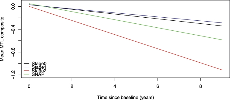
This study examined whether longitudinal MRI trajectories in medial temporal lobe (MTL) brain regions differed among groups of cognitively normal individuals defined by their cerebrospinal fluid (CSF) levels when they were first enrolled ( = 207; mean clinical follow-up = 13.3 years (max = 20 years), mean MRI follow-up = 2.4 years (max = 8 years)). We first compared atrophy rates among groups defined by CSF amyloid and phosphorylated-tau (p-tau) vs. CSF amyloid and total tau (t-tau). We also examined whether, in the presence of amyloid or tau/p-tau, the atrophy rates differed based on whether the subjects ultimately progressed to a diagnosis of mild cognitive impairment (MCI), as well as whether apolipoprotein ε4 (Apoε4) status had an impact on the longitudinal MRI trajectories. The primary finding was that when the groups were defined using CSF amyloid and p-tau, individuals with low levels of CSF amyloid and high levels of CSF p-tau (referred to as Stage 2) showed a significantly greater rate of atrophy in a composite measure of MTL volumes compared to groups defined by evidence of abnormal CSF levels in only one of the brain proteins (but not both), or no evidence of CSF abnormality. In contrast, there were no differences in rate of MTL atrophy when the groups were defined by levels of CSF amyloid and t-tau (instead of p-tau). Additionally, the rate of MTL atrophy did not differ between subjects who progressed to MCI at follow-up vs. those who remained cognitively normal when CSF levels of amyloid, t-tau, or p-tau were covaried. Lastly, the presence of an APOE ε4 genotype did not modulate the degree of MTL atrophy once baseline levels of CSF amyloid, p-tau or t-tau were accounted for. These results suggest that abnormal levels of CSF amyloid and CSF p-tau (but not t-tau) maximize the likelihood of observing significant MTL atrophy over time among individuals with normal cognition at baseline, and emphasize the importance of differentiating biomarkers that primarily reflect neurofibrillary tangle pathology (CSF p-tau) compared with biomarkers of neuronal injury (CSF t-tau).
本研究旨在探讨在认知正常个体中,当根据首次入组时的脑脊液(CSF)水平(n=207;平均临床随访时间为 13.3 年(最长 20 年),平均 MRI 随访时间为 2.4 年(最长 8 年))将其分为不同组时,内侧颞叶(MTL)脑区的纵向 MRI 轨迹是否存在差异。我们首先比较了 CSF 淀粉样蛋白和磷酸化 tau(p-tau)与 CSF 淀粉样蛋白和总 tau(t-tau)定义的组之间的萎缩率。我们还研究了在存在淀粉样蛋白或 tau/p-tau 的情况下,根据最终是否发展为轻度认知障碍(MCI)的诊断,以及载脂蛋白 E4(Apoε4)状态是否对纵向 MRI 轨迹产生影响,萎缩率是否存在差异。主要发现是,当根据 CSF 淀粉样蛋白和 p-tau 定义组时,与仅一种脑蛋白(而不是两种)的 CSF 水平异常或无 CSF 异常证据的组相比,CSF 淀粉样蛋白水平低且 CSF p-tau 水平高(称为第 2 阶段)的个体,其 MTL 容积综合测量的萎缩率显著更高。相比之下,当根据 CSF 淀粉样蛋白和 t-tau(而非 p-tau)定义组时,MTL 萎缩率没有差异。此外,当 CSF 淀粉样蛋白、t-tau 或 p-tau 水平相同时,在随访时进展为 MCI 的受试者与认知正常的受试者之间的 MTL 萎缩率没有差异。最后,在考虑基线 CSF 淀粉样蛋白、p-tau 或 t-tau 水平后,APOE ε4 基因型的存在并未调节 MTL 萎缩的程度。这些结果表明,在基线认知正常的个体中,CSF 淀粉样蛋白和 CSF p-tau(而非 t-tau)的异常水平最大程度地增加了随时间观察到显著 MTL 萎缩的可能性,并且强调了区分主要反映神经纤维缠结病理(CSF p-tau)的生物标志物与神经元损伤(CSF t-tau)生物标志物的重要性。