• 文献检索
  • 文档翻译
  • 深度研究
  • 学术资讯
  • Suppr Zotero 插件Zotero 插件
  • 邀请有礼
  • 套餐&价格
  • 历史记录
应用&插件
Suppr Zotero 插件Zotero 插件浏览器插件Mac 客户端Windows 客户端微信小程序
定价
高级版会员购买积分包购买API积分包
服务
文献检索文档翻译深度研究API 文档MCP 服务
关于我们
关于 Suppr公司介绍联系我们用户协议隐私条款
关注我们

Suppr 超能文献

核心技术专利:CN118964589B侵权必究
粤ICP备2023148730 号-1Suppr @ 2026

文献检索

告别复杂PubMed语法,用中文像聊天一样搜索,搜遍4000万医学文献。AI智能推荐,让科研检索更轻松。

立即免费搜索

文件翻译

保留排版,准确专业,支持PDF/Word/PPT等文件格式,支持 12+语言互译。

免费翻译文档

深度研究

AI帮你快速写综述,25分钟生成高质量综述,智能提取关键信息,辅助科研写作。

立即免费体验

使用具有有机硅、葡聚糖和聚乙二醇涂层的超小超顺磁性氧化铁纳米颗粒对间充质干细胞进行直接标记的效果和耐久性

Efficacy and Durability in Direct Labeling of Mesenchymal Stem Cells Using Ultrasmall Superparamagnetic Iron Oxide Nanoparticles with Organosilica, Dextran, and PEG Coatings.

作者信息

Wang Yi-Xiang J, Quercy-Jouvet Thibault, Wang Hao-Hao, Li Ak-Wai, Chak Chun-Pong, Xuan Shouhu, Shi Lin, Wang De-Feng, Lee Siu-Fung, Leung Ping-Chung, Lau Clara B S, Fung Kwok-Pui, Leung Ken Cham-Fai

机构信息

Department of Imaging and Interventional Radiology, Prince of Wales Hospital, The Chinese University of Hong Kong, Shatin, NT, Hong Kong, China.

Center of Novel Functional Molecules and Institute of Molecular Functional Materials, Department of Chemistry, The Chinese University of Hong Kong, Shatin, NT, Hong Kong, China.

出版信息

Materials (Basel). 2011 Apr 7;4(4):703-715. doi: 10.3390/ma4040703.

DOI:10.3390/ma4040703
PMID:28879947
原文链接:https://pmc.ncbi.nlm.nih.gov/articles/PMC5448517/
Abstract

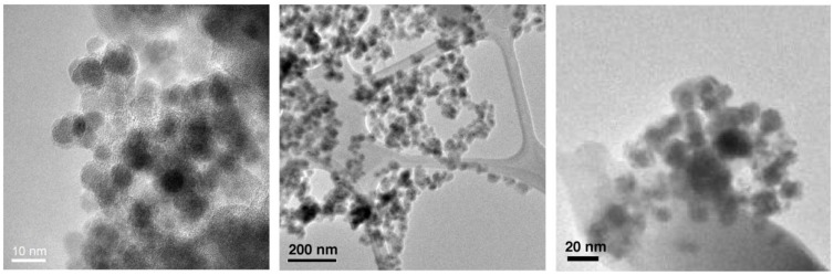
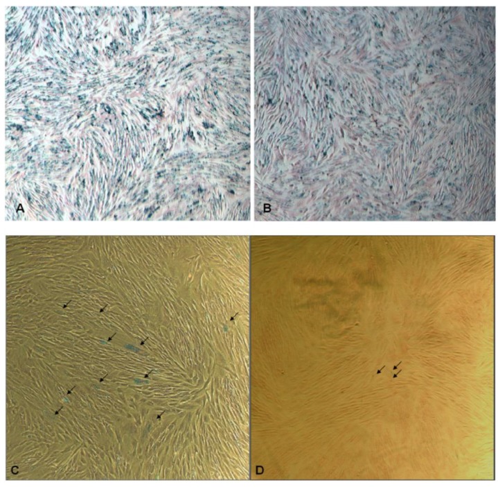
We herein report a comparative study of mesenchymal stem cell (MSC) labeling using spherical superparamagnetic iron oxide (SPIO) nanoparticles containing different coatings, namely, organosilica, dextran, and poly(ethylene glycol) (PEG). These nanomaterials possess a similar SPIO core size of 6-7 nm. Together with their coatings, the overall sizes are 10-15 nm for all SPIO@SiO₂, SPIO@dextran, and SPIO@PEG nanoparticles. These nanoparticles were investigated for their efficacies to be uptaken by rabbit bone marrow-derived MSCs without any transfecting agent. Experimentally, both SPIO@SiO₂ and SPIO@PEG nanoparticles could be successfully uptaken by MSCs while the SPIO@dextran nanoparticles demonstrated limited labeling efficiency. The labeling durability of SPIO@SiO₂ and SPIO@PEG nanoparticles in MSCs after three weeks of culture were compared by Prussian blue staining tests. SPIO@SiO₂ nanoparticles demonstrated more blue staining than SPIO@PEG nanoparticles, rendering them better materials for MSCs labeling by direct uptake when durable intracellullar retention of SPIO is desired.

摘要

我们在此报告一项关于使用含有不同涂层(即有机硅、葡聚糖和聚乙二醇(PEG))的球形超顺磁性氧化铁(SPIO)纳米颗粒对间充质干细胞(MSC)进行标记的比较研究。这些纳米材料具有相似的6 - 7纳米的SPIO核心尺寸。连同它们的涂层,所有SPIO@SiO₂、SPIO@葡聚糖和SPIO@PEG纳米颗粒的总体尺寸为10 - 15纳米。在没有任何转染剂的情况下,研究了这些纳米颗粒被兔骨髓来源的间充质干细胞摄取的效率。实验表明,SPIO@SiO₂和SPIO@PEG纳米颗粒都能被间充质干细胞成功摄取,而SPIO@葡聚糖纳米颗粒的标记效率有限。通过普鲁士蓝染色试验比较了SPIO@SiO₂和SPIO@PEG纳米颗粒在间充质干细胞中培养三周后的标记耐久性。SPIO@SiO₂纳米颗粒显示出比SPIO@PEG纳米颗粒更多的蓝色染色,当需要SPIO在细胞内持久保留时,使其成为通过直接摄取对间充质干细胞进行标记的更好材料。

https://cdn.ncbi.nlm.nih.gov/pmc/blobs/a7f6/5448517/7cf60d819936/materials-04-00703-g003.jpg
https://cdn.ncbi.nlm.nih.gov/pmc/blobs/a7f6/5448517/d038bfe6f20a/materials-04-00703-g001.jpg
https://cdn.ncbi.nlm.nih.gov/pmc/blobs/a7f6/5448517/059a5ef9bb09/materials-04-00703-g002.jpg
https://cdn.ncbi.nlm.nih.gov/pmc/blobs/a7f6/5448517/7cf60d819936/materials-04-00703-g003.jpg
https://cdn.ncbi.nlm.nih.gov/pmc/blobs/a7f6/5448517/d038bfe6f20a/materials-04-00703-g001.jpg
https://cdn.ncbi.nlm.nih.gov/pmc/blobs/a7f6/5448517/059a5ef9bb09/materials-04-00703-g002.jpg
https://cdn.ncbi.nlm.nih.gov/pmc/blobs/a7f6/5448517/7cf60d819936/materials-04-00703-g003.jpg

相似文献

1
Efficacy and Durability in Direct Labeling of Mesenchymal Stem Cells Using Ultrasmall Superparamagnetic Iron Oxide Nanoparticles with Organosilica, Dextran, and PEG Coatings.使用具有有机硅、葡聚糖和聚乙二醇涂层的超小超顺磁性氧化铁纳米颗粒对间充质干细胞进行直接标记的效果和耐久性
Materials (Basel). 2011 Apr 7;4(4):703-715. doi: 10.3390/ma4040703.
2
Enhanced cellular uptake of aminosilane-coated superparamagnetic iron oxide nanoparticles in mammalian cell lines.氨基硅烷修饰的超顺磁性氧化铁纳米颗粒在哺乳动物细胞系中的增强细胞摄取。
Int J Nanomedicine. 2012;7:953-64. doi: 10.2147/IJN.S28316. Epub 2012 Feb 21.
3
Durable mesenchymal stem cell labelling by using polyhedral superparamagnetic iron oxide nanoparticles.多面体超顺磁性氧化铁纳米粒子对间充质干细胞的长期标记。
Chemistry. 2009 Nov 16;15(45):12417-25. doi: 10.1002/chem.200901548.
4
Polyethylene glycol–coated and folic acid–conjugated superparamagnetic iron oxide nanoparticles聚乙二醇包覆且叶酸偶联的超顺磁性氧化铁纳米颗粒
5
Poly(,-dimethylacrylamide)-coated maghemite nanoparticles for labeling and tracking mesenchymal stem cells用于标记和追踪间充质干细胞的聚(N,N-二甲基丙烯酰胺)包覆磁赤铁矿纳米颗粒
6
Thiol-modified poly(ethylene glycol)-conjugated gold/superparamagnetic iron oxide nanoparticles硫醇修饰的聚乙二醇共轭金/超顺磁性氧化铁纳米颗粒
7
Amine-modified silica-coated polyhedral superparamagnetic iron oxide nanoparticle–labeled rabbit bone marrow–derived mesenchymal stem cells胺修饰的二氧化硅包覆多面体超顺磁性氧化铁纳米颗粒标记的兔骨髓间充质干细胞
8
Magnetic resonance and photoacoustic imaging of brain tumor mediated by mesenchymal stem cell labeled with multifunctional nanoparticle introduced via carotid artery injection.经颈动脉注射导入多功能纳米颗粒标记的间充质干细胞介导的脑肿瘤磁共振和光声成像。
Nanotechnology. 2018 Apr 20;29(16):165101. doi: 10.1088/1361-6528/aaaf16.
9
Evaluation of umbilical cord mesenchymal stem cell labeling with superparamagnetic iron oxide nanoparticles coated with dextran and complexed with Poly-L-lysine.用葡聚糖包被并与聚-L-赖氨酸络合的超顺磁性氧化铁纳米颗粒标记脐带间充质干细胞的评估。
Einstein (Sao Paulo). 2012 Apr-Jun;10(2):180-8. doi: 10.1590/s1679-45082012000200011.
10
FluidMAG iron nanoparticle-labeled mesenchymal stem cells for tracking cell homing to tumors用于追踪细胞归巢至肿瘤的流体磁体铁纳米颗粒标记间充质干细胞

引用本文的文献

1
Synthesis and Characterization of Citric Acid-Modified Iron Oxide Nanoparticles Prepared with Electrohydraulic Discharge Treatment.用电液压放电处理制备的柠檬酸改性氧化铁纳米颗粒的合成与表征
Materials (Basel). 2023 Jan 12;16(2):746. doi: 10.3390/ma16020746.
2
Citrate-Coated Magnetic Polyethyleneimine Composites for Plasmid DNA Delivery into Glioblastoma.用于将质粒DNA递送至胶质母细胞瘤的柠檬酸盐包覆磁性聚乙烯亚胺复合材料
Polymers (Basel). 2021 Jul 6;13(14):2228. doi: 10.3390/polym13142228.
3
A brief review of cytotoxicity of nanoparticles on mesenchymal stem cells in regenerative medicine.

本文引用的文献

1
Color quantification for evaluation of stained tissues.用于评估染色组织的颜色量化。
Cytometry A. 2011 Apr;79(4):311-6. doi: 10.1002/cyto.a.21037. Epub 2011 Mar 8.
2
Low-intensity pulsed ultrasound increases cellular uptake of superparamagnetic iron oxide nanomaterial: results from human osteosarcoma cell line U2OS.低强度脉冲超声增强超顺磁性氧化铁纳米材料的细胞摄取:来自人骨肉瘤细胞系 U2OS 的结果。
J Magn Reson Imaging. 2010 Jun;31(6):1508-13. doi: 10.1002/jmri.22173.
3
Durable mesenchymal stem cell labelling by using polyhedral superparamagnetic iron oxide nanoparticles.
纳米颗粒对再生医学中间充质干细胞细胞毒性的简要综述。
Int J Nanomedicine. 2019 May 24;14:3875-3892. doi: 10.2147/IJN.S205574. eCollection 2019.
4
Evaluation of biocompatible alginate- and deferoxamine-coated ternary composites for magnetic resonance imaging and gene delivery into glioblastoma cells.用于磁共振成像以及将基因递送至胶质母细胞瘤细胞的生物相容性藻酸盐和去铁胺包被的三元复合材料的评估。
Quant Imaging Med Surg. 2015 Jun;5(3):382-91. doi: 10.3978/j.issn.2223-4292.2015.03.12.
5
Nanoparticle-encapsulated chlorhexidine against oral bacterial biofilms.纳米颗粒包裹的洗必泰对口腔细菌生物膜的作用
PLoS One. 2014 Aug 29;9(8):e103234. doi: 10.1371/journal.pone.0103234. eCollection 2014.
6
Ternary hybrid nanocomposites for gene delivery and magnetic resonance imaging of hepatocellular carcinoma cells.用于肝细胞癌基因递送和磁共振成像的三元杂化纳米复合材料。
Quant Imaging Med Surg. 2013 Dec;3(6):302-7. doi: 10.3978/j.issn.2223-4292.2013.12.05.
7
Superparamagnetic iron oxide nanoparticles as MRI contrast agents for non-invasive stem cell labeling and tracking.超顺磁性氧化铁纳米颗粒作为 MRI 对比剂用于非侵入性干细胞标记和示踪。
Theranostics. 2013 Jul 31;3(8):595-615. doi: 10.7150/thno.5366. eCollection 2013.
8
Recent advances in superparamagnetic iron oxide nanoparticles for cellular imaging and targeted therapy research.超顺磁性氧化铁纳米粒子在细胞成像和靶向治疗研究中的最新进展。
Curr Pharm Des. 2013;19(37):6575-93. doi: 10.2174/1381612811319370003.
9
Preparation and characterization of ferrofluid stabilized with biocompatible chitosan and dextran sulfate hybrid biopolymer as a potential magnetic resonance imaging (MRI) T2 contrast agent.用生物相容性壳聚糖和葡聚糖硫酸酯杂化生物聚合物稳定的铁磁流体的制备和特性,作为一种潜在的磁共振成像(MRI)T2 对比剂。
Mar Drugs. 2012 Oct 29;10(11):2403-14. doi: 10.3390/md10112403.
多面体超顺磁性氧化铁纳米粒子对间充质干细胞的长期标记。
Chemistry. 2009 Nov 16;15(45):12417-25. doi: 10.1002/chem.200901548.
4
Biological and magnetic contrast evaluation of shape-selective Mn-Fe nanowires.形状选择性锰铁纳米线的生物和磁性对比评估
IEEE Trans Nanobioscience. 2009 Jun;8(2):192-8. doi: 10.1109/TNB.2009.2021521. Epub 2009 May 8.
5
MR tracking of stem cells in living recipients.活体受体中干细胞的磁共振追踪
Methods Mol Biol. 2009;549:197-215. doi: 10.1007/978-1-60327-931-4_14.
6
Systematic surface engineering of magnetic nanoworms for in vivo tumor targeting.用于体内肿瘤靶向的磁性纳米蠕虫的系统表面工程。
Small. 2009 Mar;5(6):694-700. doi: 10.1002/smll.200801789.
7
Physical approaches to biomaterial design.生物材料设计的物理方法。
Nat Mater. 2009 Jan;8(1):15-23. doi: 10.1038/nmat2344.
8
Comment on Jing et al. original article "In vivo MR imaging tracking of magnetic iron oxide nanoparticle labeled, engineered, autologous bone marrow mesenchymal stem cells following intra-articular injection".对景等人的原创文章《关节腔内注射后磁性氧化铁纳米颗粒标记的工程化自体骨髓间充质干细胞的体内磁共振成像追踪》的评论
Joint Bone Spine. 2009 Jan;76(1):118-9; author reply 119-20. doi: 10.1016/j.jbspin.2008.09.008. Epub 2008 Dec 17.
9
Pitfalls in employing superparamagnetic iron oxide particles for stem cell labelling and in vivo MRI tracking.使用超顺磁性氧化铁颗粒进行干细胞标记和体内MRI追踪的陷阱。
Br J Radiol. 2008 Dec;81(972):987-8. doi: 10.1259/bjr/55991430.
10
Magnetic iron oxide nanoparticles: synthesis, stabilization, vectorization, physicochemical characterizations, and biological applications.磁性氧化铁纳米颗粒:合成、稳定化、载体化、物理化学表征及生物学应用。
Chem Rev. 2008 Jun;108(6):2064-110. doi: 10.1021/cr068445e.