Davenport Kristen A, Hoover Clare E, Bian Jifeng, Telling Glenn C, Mathiason Candace K, Hoover Edward A
Prion Research Center, Microbiology, Immunology and Pathology Department, Colorado State University, Fort Collins, Colorado, United States of America.
PLoS One. 2017 Sep 7;12(9):e0183927. doi: 10.1371/journal.pone.0183927. eCollection 2017.
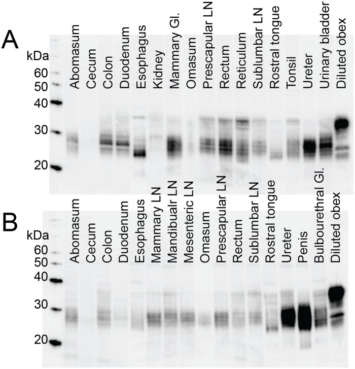
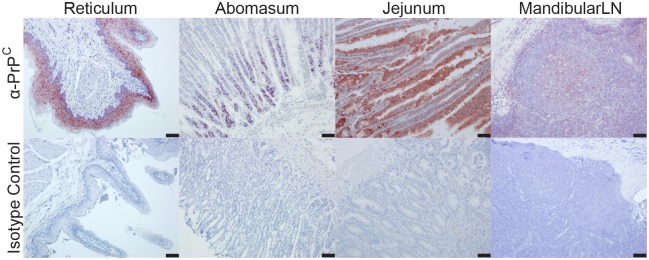
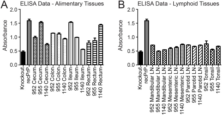
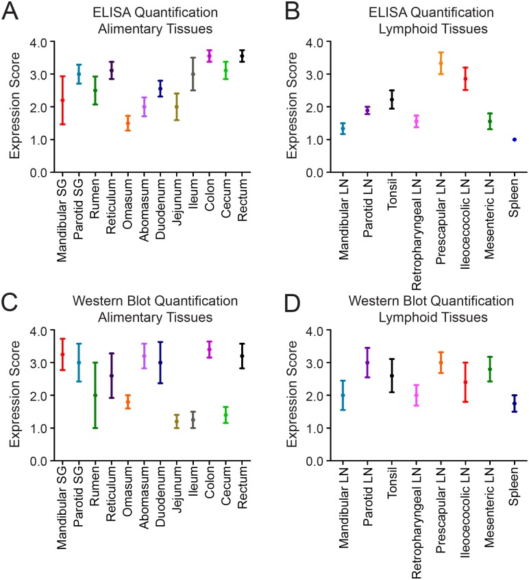
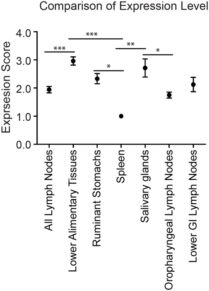
The agent responsible for prion diseases is a misfolded form of a normal protein (PrPC). The prion hypothesis stipulates that PrPC must be present for the disease to manifest. Cervid populations across the world are infected with chronic wasting disease, a horizontally-transmissible prion disease that is likely spread via oral exposure to infectious prions (PrPCWD). Though PrPCWD has been identified in many tissues, there has been little effort to characterize the overall PrPC expression in cervids and its relationship to PrPCWD accumulation. We used immunohistochemistry (IHC), western blot and enzyme-linked immunosorbent assay to describe PrPC expression in naïve white-tailed deer. We used real-time, quaking-induced conversion (RT-QuIC) to detect prion seeding activity in CWD-infected deer. We assessed tissues comprising the alimentary tract, alimentary-associated lymphoid tissue and systemic lymphoid tissue from 5 naïve deer. PrPC was expressed in all tissues, though expression was often very low compared to the level in the CNS. IHC identified specific cell types wherein PrPC expression is very high. To compare the distribution of PrPC to PrPCWD, we examined 5 deer with advanced CWD infection. Using RT-QuIC, we detected prion seeding activity in all 21 tissues. In 3 subclinical deer sacrificed 4 months post-inoculation, we detected PrPCWD consistently in alimentary-associated lymphoid tissue, irregularly in alimentary tract tissues, and not at all in the brain. Contrary to our hypothesis that PrPC levels dictate prion accumulation, PrPC expression was higher in the lower gastrointestinal tissues than in the alimentary-associated lymphoid system and was higher in salivary glands than in the oropharyngeal lymphoid tissue. These data suggest that PrPC expression is not the sole driver of prion accumulation and that alimentary tract tissues accumulate prions before centrifugal spread from the brain occurs.
导致朊病毒疾病的病原体是一种正常蛋白质(PrPC)的错误折叠形式。朊病毒假说规定,疾病的显现必须有PrPC的存在。世界各地的鹿群感染了慢性消耗病,这是一种可横向传播的朊病毒疾病,可能通过口腔接触传染性朊病毒(PrPCWD)传播。尽管在许多组织中都已鉴定出PrPCWD,但在鹿中对整体PrPC表达及其与PrPCWD积累的关系进行表征的工作却很少。我们使用免疫组织化学(IHC)、蛋白质印迹法和酶联免疫吸附测定来描述未感染的白尾鹿中的PrPC表达。我们使用实时、颤抖诱导转化(RT-QuIC)来检测慢性消耗病感染鹿中的朊病毒播种活性。我们评估了5只未感染鹿的包括消化道、消化道相关淋巴组织和全身淋巴组织的组织。PrPC在所有组织中均有表达,尽管与中枢神经系统中的水平相比,其表达通常非常低。免疫组织化学鉴定了PrPC表达非常高的特定细胞类型。为了比较PrPC与PrPCWD的分布,我们检查了5只患有晚期慢性消耗病感染的鹿。使用RT-QuIC,我们在所有21种组织中均检测到了朊病毒播种活性。在接种后4个月处死的3只亚临床鹿中,我们在消化道相关淋巴组织中始终检测到PrPCWD,在消化道组织中检测到的情况不规则,而在脑中则完全未检测到。与我们认为PrPC水平决定朊病毒积累的假设相反,PrPC在下消化道组织中的表达高于消化道相关淋巴系统,在唾液腺中的表达高于口咽淋巴组织。这些数据表明,PrPC表达不是朊病毒积累的唯一驱动因素,并且在从脑发生离心扩散之前,消化道组织就积累了朊病毒。