Advanced Imaging Research Center, University of Texas Southwestern Medical Center at Dallas, Dallas, TX 75390, USA; Organ Transplantation Center, The First Affiliated Hospital, Sun Yat-sen University, Guangzhou, PR China.
Advanced Imaging Research Center, University of Texas Southwestern Medical Center at Dallas, Dallas, TX 75390, USA.
Free Radic Biol Med. 2017 Nov;112:597-607. doi: 10.1016/j.freeradbiomed.2017.09.002. Epub 2017 Sep 8.
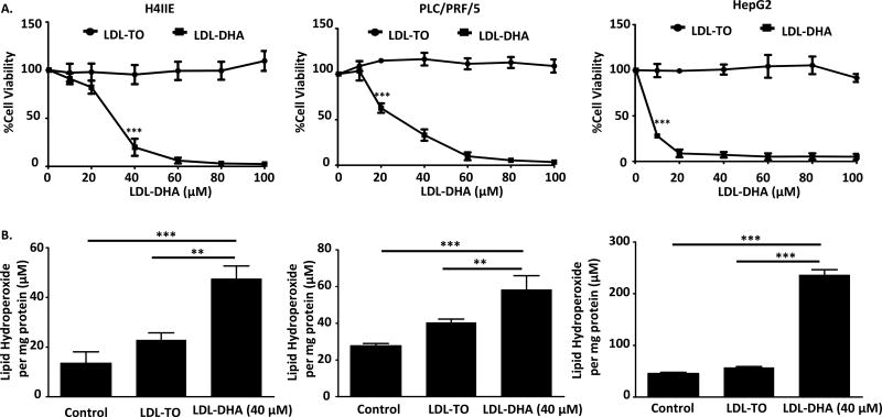
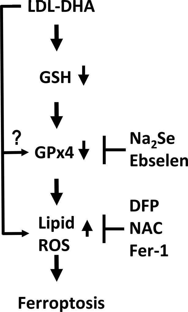
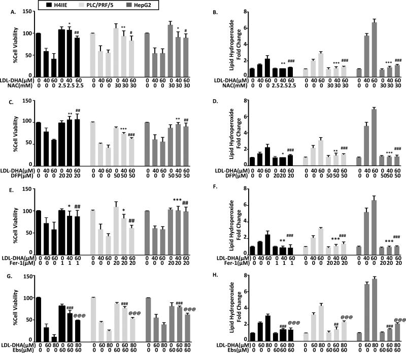
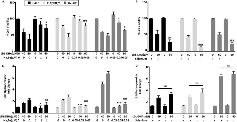
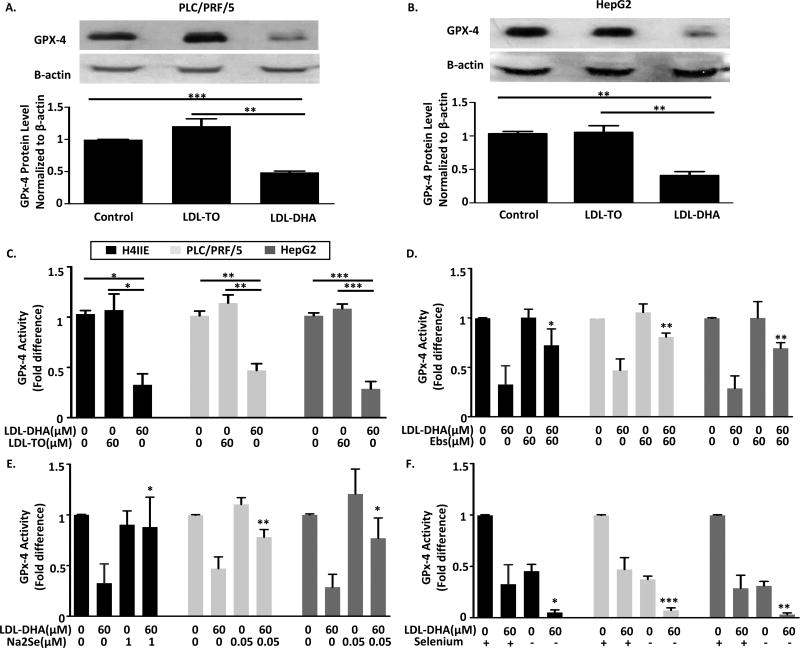
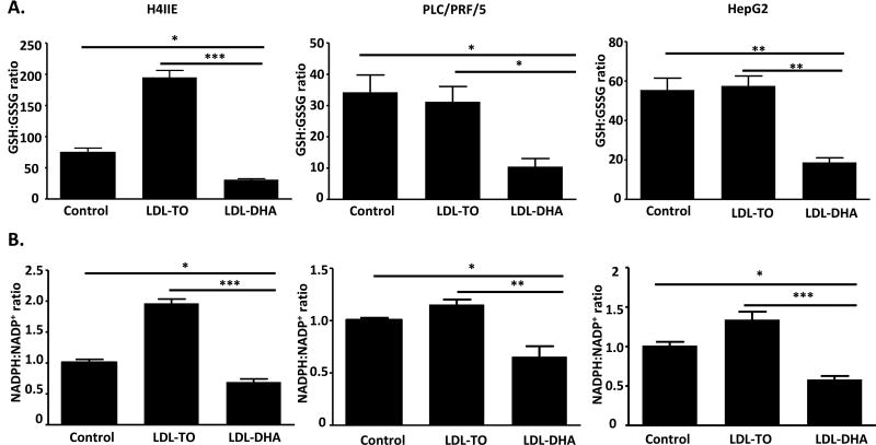
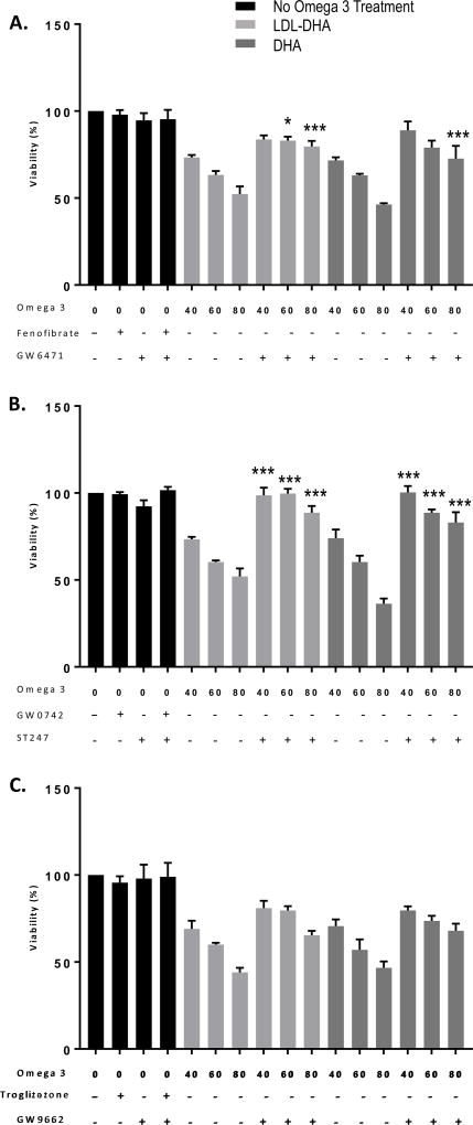
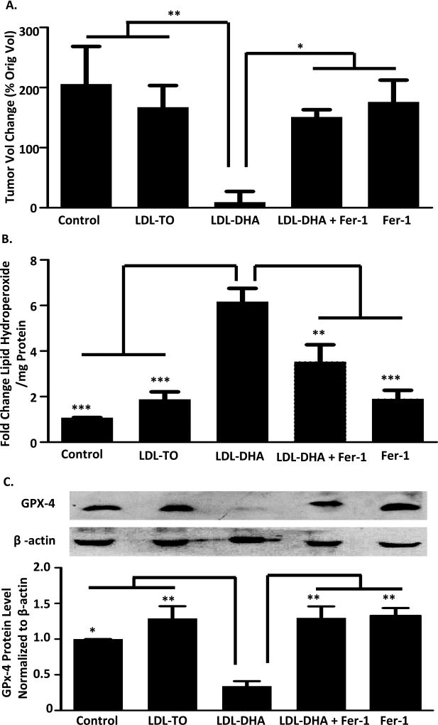
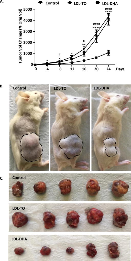
Low-density lipoprotein nanoparticles reconstituted with the natural omega-3 fatty acid, docosahexaenoic acid (LDL-DHA), have been reported to selectively kill hepatoma cells and reduce the growth of orthotopic liver tumors in the rat. To date, little is known about the cell death pathways by which LDL-DHA nanoparticles kill tumor cells. Here we show that the LDL-DHA nanoparticles are cytotoxic to both rat hepatoma and human hepatocellular carcinoma (HCC) cell lines. Following LDL-DHA treatment both rat and human HCC cells experience pronounced lipid peroxidation, depletion of glutathione and inactivation of the lipid antioxidant glutathione peroxidase-4 (GPX4) prior to cell death. Inhibitor studies revealed that the treated HCC cells die independent of apoptotic, necroptotic or autophagic pathways, but require the presence of cellular iron. These hallmark features are consistent and were later confirmed to reflect ferroptosis, a novel form of nonapoptotic iron-dependent cell death. In keeping with the mechanisms of ferroptosis cell death, GPX4 was also found to be a central regulator of LDL-DHA induced tumor cell killing. We also investigated the effects of LDL-DHA treatments in mice bearing human HCC tumor xenografts. Intratumoral injections of LDL-DHA severely inhibited the growth of HCC xenografts long term. Consistent with our in vitro findings, the LDL-DHA treated HCC tumors experienced ferroptotic cell death characterized by increased levels of tissue lipid hydroperoxides and suppression of GPX4 expression.
LDL-DHA induces cell death in HCC cells through the ferroptosis pathway, this represents a novel molecular mechanism of anticancer activity for LDL-DHA nanoparticles.
用天然 ω-3 脂肪酸二十二碳六烯酸(DHA)重建的低密度脂蛋白纳米颗粒已被报道可选择性杀死肝癌细胞并减少大鼠原位肝肿瘤的生长。迄今为止,人们对 LDL-DHA 纳米颗粒杀死肿瘤细胞的细胞死亡途径知之甚少。在这里,我们表明 LDL-DHA 纳米颗粒对大鼠肝癌和人肝癌细胞系均具有细胞毒性。在 LDL-DHA 处理后,大鼠和人 HCC 细胞均经历明显的脂质过氧化,谷胱甘肽耗竭和脂质抗氧化剂谷胱甘肽过氧化物酶-4(GPX4)失活,然后发生细胞死亡。抑制剂研究表明,经处理的 HCC 细胞死亡不依赖于凋亡,坏死或自噬途径,但需要存在细胞内铁。这些标志性特征是一致的,并随后被证实反映了铁死亡,这是一种新型的非凋亡性铁依赖性细胞死亡形式。与铁死亡细胞死亡的机制一致,GPX4 也被发现是 LDL-DHA 诱导的肿瘤细胞杀伤的中央调节剂。我们还研究了 LDL-DHA 处理在携带人 HCC 肿瘤异种移植物的小鼠中的作用。LDL-DHA 的肿瘤内注射可长期严重抑制 HCC 异种移植物的生长。与我们的体外发现一致,LDL-DHA 处理的 HCC 肿瘤经历了铁死亡,其特征是组织脂质过氧化物水平升高和 GPX4 表达抑制。
LDL-DHA 通过铁死亡途径诱导 HCC 细胞死亡,这代表了 LDL-DHA 纳米颗粒的抗癌活性的新分子机制。