Department of Radiology, Leiden University Medical Center , Leiden 2333 ZA, Netherlands.
Department of Neurobiology, Weizmann Institute of Science , Rehovot 7610001, Israel.
ACS Appl Mater Interfaces. 2017 Oct 11;9(40):34618-34624. doi: 10.1021/acsami.7b06949. Epub 2017 Sep 26.
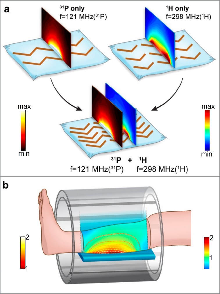
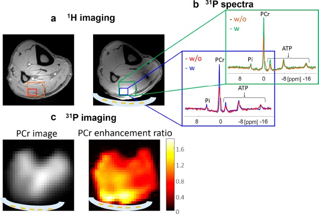
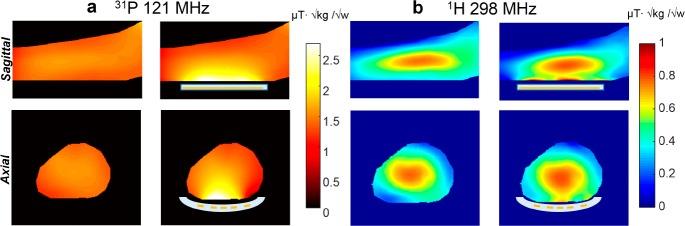
Magnetic resonance imaging and spectroscopy (MRI and MRS) are both widely used techniques in medical diagnostics and research. One of the major thrusts in recent years has been the introduction of ultrahigh-field magnets in order to boost the sensitivity. Several MRI studies have examined further potential improvements in sensitivity using metamaterials, focusing on single frequency applications. However, metamaterials have yet to reach a level that is practical for routine MRI use. In this work, we explore a new metamaterial implementation for MRI, a dual-nuclei resonant structure, which can be used for both proton and heteronuclear magnetic resonance. Our approach combines two configurations, one based on a set of electric dipoles for the low frequency band, and the second based on a set of magnetic dipoles for the high frequency band. We focus on the implementation of a dual-nuclei metamaterial for phosphorus and proton imaging and spectroscopy at an ultrahigh-field strength of 7 T. In vivo scans using this flexible and compact structure show that it locally enhances both the phosphorus and proton transmit and receive sensitivities.
磁共振成象和波谱学(MRI 和 MRS)都是医学诊断和研究中广泛应用的技术。近年来的一个主要发展趋势是引入超高磁场磁铁,以提高灵敏度。一些 MRI 研究使用超材料进一步探讨了提高灵敏度的潜力,重点是单频应用。然而,超材料尚未达到实际用于常规 MRI 的水平。在这项工作中,我们探索了一种新的 MRI 超材料实现方法,即双核共振结构,可用于质子和异核磁共振。我们的方法结合了两种配置,一种基于低频带的一组电偶极子,另一种基于高频带的一组磁偶极子。我们专注于在 7T 的超高场强下实现磷和质子成像和波谱学的双核超材料。使用这种灵活紧凑结构的体内扫描表明,它局部增强了磷和质子的发射和接收灵敏度。