• 文献检索
  • 文档翻译
  • 深度研究
  • 学术资讯
  • Suppr Zotero 插件Zotero 插件
  • 邀请有礼
  • 套餐&价格
  • 历史记录
应用&插件
Suppr Zotero 插件Zotero 插件浏览器插件Mac 客户端Windows 客户端微信小程序
定价
高级版会员购买积分包购买API积分包
服务
文献检索文档翻译深度研究API 文档MCP 服务
关于我们
关于 Suppr公司介绍联系我们用户协议隐私条款
关注我们

Suppr 超能文献

核心技术专利:CN118964589B侵权必究
粤ICP备2023148730 号-1Suppr @ 2026

文献检索

告别复杂PubMed语法,用中文像聊天一样搜索,搜遍4000万医学文献。AI智能推荐,让科研检索更轻松。

立即免费搜索

文件翻译

保留排版,准确专业,支持PDF/Word/PPT等文件格式,支持 12+语言互译。

免费翻译文档

深度研究

AI帮你快速写综述,25分钟生成高质量综述,智能提取关键信息,辅助科研写作。

立即免费体验

基于移动设备的疟疾感染薄血涂片分析:自动鉴定物种和生活史阶段。

Mobile-Based Analysis of Malaria-Infected Thin Blood Smears: Automated Species and Life Cycle Stage Determination.

机构信息

Fraunhofer Portugal AICOS, Rua Alfredo Allen 455/461, 4200-135 Porto, Portugal.

Instituto Nacional de Saúde Dr. Ricardo Jorge, Rua Alexandre Herculano 321, 4000-055 Porto, Portugal.

出版信息

Sensors (Basel). 2017 Sep 21;17(10):2167. doi: 10.3390/s17102167.

DOI:10.3390/s17102167
PMID:28934170
原文链接:https://pmc.ncbi.nlm.nih.gov/articles/PMC5677014/
Abstract

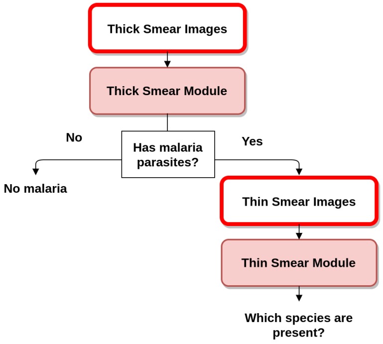
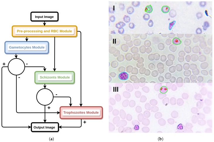
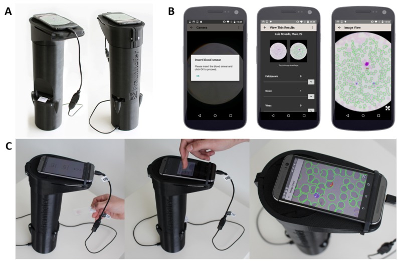
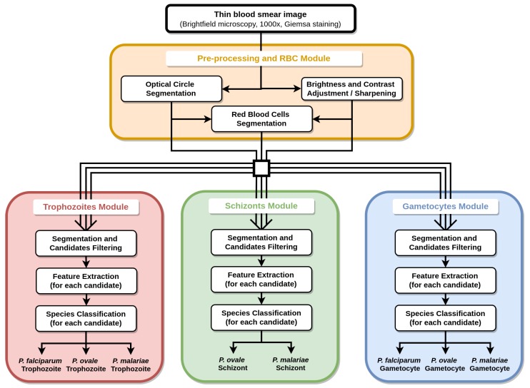
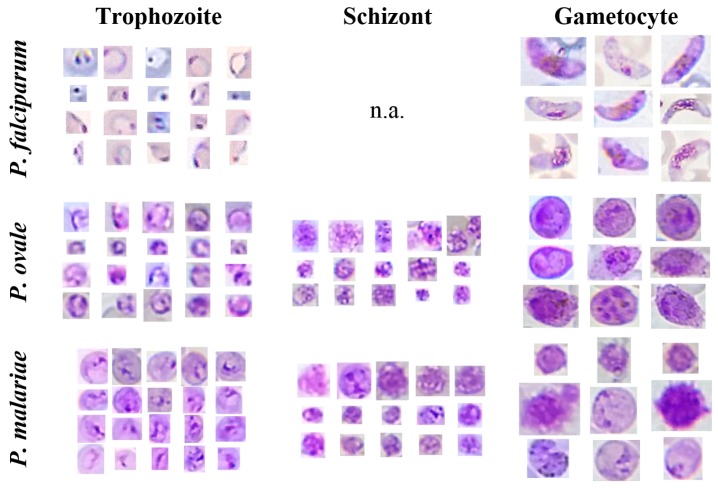
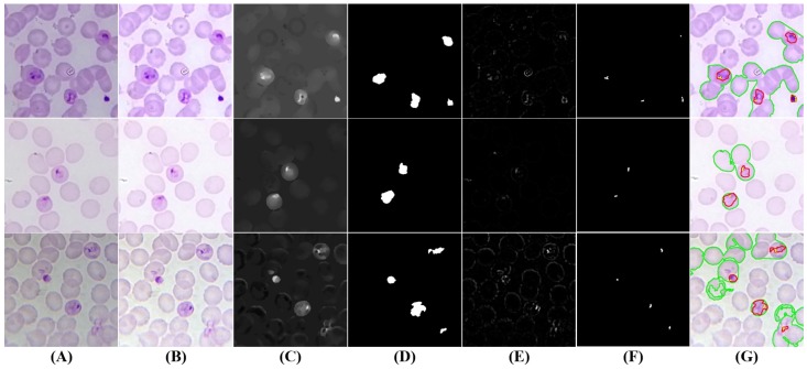
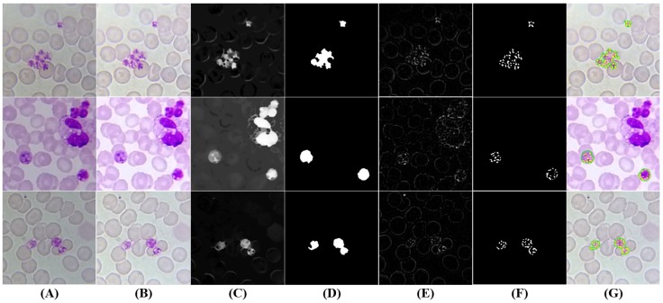
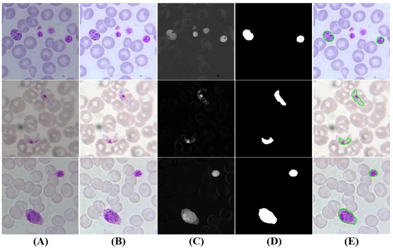
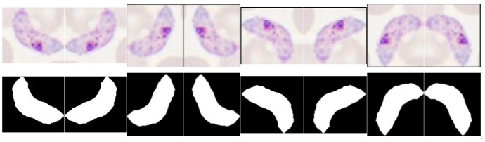
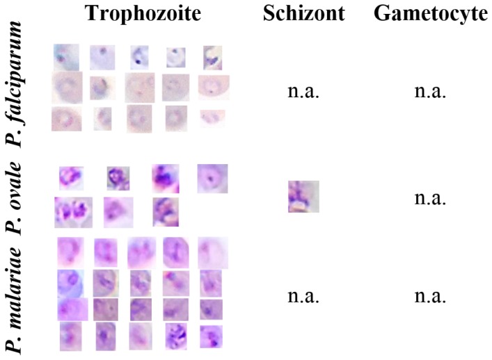
Microscopy examination has been the pillar of malaria diagnosis, being the recommended procedure when its quality can be maintained. However, the need for trained personnel and adequate equipment limits its availability and accessibility in malaria-endemic areas. Rapid, accurate, accessible diagnostic tools are increasingly required, as malaria control programs extend parasite-based diagnosis and the prevalence decreases. This paper presents an image processing and analysis methodology using supervised classification to assess the presence of malaria parasites and determine the species and life cycle stage in Giemsa-stained thin blood smears. The main differentiation factor is the usage of microscopic images exclusively acquired with low cost and accessible tools such as smartphones, a dataset of 566 images manually annotated by an experienced parasilogist being used. Eight different species-stage combinations were considered in this work, with an automatic detection performance ranging from 73.9% to 96.2% in terms of sensitivity and from 92.6% to 99.3% in terms of specificity. These promising results attest to the potential of using this approach as a valid alternative to conventional microscopy examination, with comparable detection performances and acceptable computational times.

摘要

显微镜检查一直是疟疾诊断的支柱,当能够保证其质量时,它是推荐的程序。然而,由于需要经过培训的人员和足够的设备,其在疟疾流行地区的可用性和可及性受到限制。随着疟疾控制项目扩展寄生虫诊断,并且患病率下降,快速、准确、易于获取的诊断工具越来越受到需求。本文提出了一种使用监督分类的图像处理和分析方法,用于评估在吉姆萨染色的薄血涂片上是否存在疟原虫,并确定其种类和生活史阶段。主要的区分因素是仅使用低成本和易于获取的工具(如智能手机)获取的显微镜图像,使用了一个由经验丰富的寄生虫学家手动注释的 566 张图像数据集。在这项工作中考虑了八种不同的种-期组合,在灵敏度方面,自动检测性能的范围为 73.9%至 96.2%,在特异性方面,自动检测性能的范围为 92.6%至 99.3%。这些有希望的结果证明了使用这种方法作为传统显微镜检查的有效替代方法的潜力,其检测性能相当,计算时间也可接受。

https://cdn.ncbi.nlm.nih.gov/pmc/blobs/9c86/5677014/3b29a8c8919f/sensors-17-02167-g014.jpg
https://cdn.ncbi.nlm.nih.gov/pmc/blobs/9c86/5677014/fa5b8ac062a7/sensors-17-02167-g001.jpg
https://cdn.ncbi.nlm.nih.gov/pmc/blobs/9c86/5677014/640a4ad63bca/sensors-17-02167-g002.jpg
https://cdn.ncbi.nlm.nih.gov/pmc/blobs/9c86/5677014/b2252633b485/sensors-17-02167-g003.jpg
https://cdn.ncbi.nlm.nih.gov/pmc/blobs/9c86/5677014/f302392640bb/sensors-17-02167-g004.jpg
https://cdn.ncbi.nlm.nih.gov/pmc/blobs/9c86/5677014/f7d36e65d094/sensors-17-02167-g005.jpg
https://cdn.ncbi.nlm.nih.gov/pmc/blobs/9c86/5677014/cc010fc1845c/sensors-17-02167-g006.jpg
https://cdn.ncbi.nlm.nih.gov/pmc/blobs/9c86/5677014/e1aa7e6d7a03/sensors-17-02167-g007.jpg
https://cdn.ncbi.nlm.nih.gov/pmc/blobs/9c86/5677014/25439a3a2a74/sensors-17-02167-g008.jpg
https://cdn.ncbi.nlm.nih.gov/pmc/blobs/9c86/5677014/590b39a7ef4e/sensors-17-02167-g009.jpg
https://cdn.ncbi.nlm.nih.gov/pmc/blobs/9c86/5677014/564979795c78/sensors-17-02167-g010.jpg
https://cdn.ncbi.nlm.nih.gov/pmc/blobs/9c86/5677014/1c7ab20a392f/sensors-17-02167-g011.jpg
https://cdn.ncbi.nlm.nih.gov/pmc/blobs/9c86/5677014/aeeadeea8360/sensors-17-02167-g012.jpg
https://cdn.ncbi.nlm.nih.gov/pmc/blobs/9c86/5677014/03d188560a2e/sensors-17-02167-g013.jpg
https://cdn.ncbi.nlm.nih.gov/pmc/blobs/9c86/5677014/3b29a8c8919f/sensors-17-02167-g014.jpg

相似文献

1
Mobile-Based Analysis of Malaria-Infected Thin Blood Smears: Automated Species and Life Cycle Stage Determination.基于移动设备的疟疾感染薄血涂片分析:自动鉴定物种和生活史阶段。
Sensors (Basel). 2017 Sep 21;17(10):2167. doi: 10.3390/s17102167.
2
An automatic device for detection and classification of malaria parasite species in thick blood film.一种用于在厚血膜中检测和分类疟原虫种类的自动设备。
BMC Bioinformatics. 2012;13 Suppl 17(Suppl 17):S18. doi: 10.1186/1471-2105-13-S17-S18. Epub 2012 Dec 13.
3
Plasmodium life cycle stage classification based quantification of malaria parasitaemia in thin blood smears.基于疟原虫生命周期阶段分类的薄血涂片疟疾寄生虫血症定量分析。
Microsc Res Tech. 2019 Mar;82(3):283-295. doi: 10.1002/jemt.23170. Epub 2018 Dec 21.
4
Automated system for characterization and classification of malaria-infected stages using light microscopic images of thin blood smears.使用薄血涂片的光学显微镜图像对疟疾感染阶段进行表征和分类的自动化系统。
J Microsc. 2015 Mar;257(3):238-52. doi: 10.1111/jmi.12206. Epub 2014 Dec 18.
5
Computational microscopic imaging for malaria parasite detection: a systematic review.用于疟原虫检测的计算显微成像:一项系统综述。
J Microsc. 2015 Oct;260(1):1-19. doi: 10.1111/jmi.12270. Epub 2015 Jun 5.
6
[Advances in automatic detection technology for images of thin blood film of malaria parasite].[疟原虫薄血膜图像自动检测技术的进展]
Zhongguo Xue Xi Chong Bing Fang Zhi Za Zhi. 2017 May 5;29(3):388-392. doi: 10.16250/j.32.1374.2017015.
7
Detection and stage classification of Plasmodium falciparum from images of Giemsa stained thin blood films using random forest classifiers.利用随机森林分类器从吉姆萨染色薄血涂片图像中检测和分期疟原虫。
Diagn Pathol. 2020 Oct 23;15(1):130. doi: 10.1186/s13000-020-01040-9.
8
Automated image processing method for the diagnosis and classification of malaria on thin blood smears.用于薄血涂片上疟疾诊断和分类的自动图像处理方法。
Med Biol Eng Comput. 2006 May;44(5):427-36. doi: 10.1007/s11517-006-0044-2. Epub 2006 Apr 8.
9
Automatic diagnosis of malaria based on complete circle-ellipse fitting search algorithm.基于完整圆-椭圆拟合搜索算法的疟疾自动诊断。
J Microsc. 2013 Dec;252(3):189-203. doi: 10.1111/jmi.12081. Epub 2013 Sep 19.
10
The diagnosis of malaria and identification of plasmodium species by polymerase chain reaction in Turkey.土耳其通过聚合酶链反应进行疟疾诊断及疟原虫种类鉴定
Parasitol Int. 2007 Sep;56(3):217-20. doi: 10.1016/j.parint.2007.03.001. Epub 2007 Mar 12.

引用本文的文献

1
A Robust Malaria Cell Detection Framework Using Adaptive and Atrous Convolution-Based Recurrent Mobilenetv2 with Trans-MobileUNet + + -Based Abnormality Segmentation.一种基于自适应空洞卷积循环MobileNetv2和基于Trans-MobileUNet++的异常分割的稳健疟疾细胞检测框架。
J Imaging Inform Med. 2024 Dec 4. doi: 10.1007/s10278-024-01311-7.
2
Advances in Portable Optical Microscopy Using Cloud Technologies and Artificial Intelligence for Medical Applications.利用云技术和人工智能的便携式光学显微镜的进展及其在医学中的应用。
Sensors (Basel). 2024 Oct 17;24(20):6682. doi: 10.3390/s24206682.
3
The Impact of Artificial Intelligence on Microbial Diagnosis.

本文引用的文献

1
A portable image-based cytometer for rapid malaria detection and quantification.一种用于快速疟疾检测和定量的便携式基于图像的细胞仪。
PLoS One. 2017 Jun 8;12(6):e0179161. doi: 10.1371/journal.pone.0179161. eCollection 2017.
2
Unsupervised malaria parasite detection based on phase spectrum.基于相位谱的无监督疟原虫检测
Annu Int Conf IEEE Eng Med Biol Soc. 2011;2011:7997-8000. doi: 10.1109/IEMBS.2011.6091972.
人工智能对微生物诊断的影响。
Microorganisms. 2024 May 23;12(6):1051. doi: 10.3390/microorganisms12061051.
4
Development of a low-cost robotized 3D-prototype for automated optical microscopy diagnosis: An open-source system.开发低成本的机器人化 3D 原型用于自动化光学显微镜诊断:一个开源系统。
PLoS One. 2024 Jun 21;19(6):e0304085. doi: 10.1371/journal.pone.0304085. eCollection 2024.
5
An Efficient and Effective Framework for Intestinal Parasite Egg Detection Using YOLOv5.一种使用YOLOv5进行肠道寄生虫卵检测的高效框架。
Diagnostics (Basel). 2023 Sep 18;13(18):2978. doi: 10.3390/diagnostics13182978.
6
Deep Learning-Based Cell Detection and Extraction in Thin Blood Smears for Malaria Diagnosis.基于深度学习的薄血涂片细胞检测与提取用于疟疾诊断
IEEE Appl Imag Pattern Recognit Workshop. 2021 Apr 26;2021:9762109. doi: 10.1109/AIPR52630.2021.9762109.
7
Advances and challenges in automated malaria diagnosis using digital microscopy imaging with artificial intelligence tools: A review.使用人工智能工具的数字显微镜成像技术在疟疾自动诊断中的进展与挑战:综述
Front Microbiol. 2022 Nov 15;13:1006659. doi: 10.3389/fmicb.2022.1006659. eCollection 2022.
8
Innovations in spp diagnosis on diverse detection platforms.不同检测平台上物种诊断的创新。
3 Biotech. 2021 Dec;11(12):505. doi: 10.1007/s13205-021-03054-6. Epub 2021 Nov 22.
9
Systematic Review and Meta-Analysis of the Diagnostic Accuracy of Mobile-Linked Point-of-Care Diagnostics in Sub-Saharan Africa.撒哈拉以南非洲地区移动连接即时诊断诊断准确性的系统评价与荟萃分析
Diagnostics (Basel). 2021 Jun 12;11(6):1081. doi: 10.3390/diagnostics11061081.
10
Automated phenotyping of mosquito larvae enables high-throughput screening for novel larvicides and offers potential for smartphone-based detection of larval insecticide resistance.蚊子幼虫的自动表型分析可实现高通量筛选新型幼虫杀虫剂,并为基于智能手机的幼虫抗药性检测提供了潜力。
PLoS Negl Trop Dis. 2021 Jun 3;15(6):e0008639. doi: 10.1371/journal.pntd.0008639. eCollection 2021 Jun.