• 文献检索
  • 文档翻译
  • 深度研究
  • 学术资讯
  • Suppr Zotero 插件Zotero 插件
  • 邀请有礼
  • 套餐&价格
  • 历史记录
应用&插件
Suppr Zotero 插件Zotero 插件浏览器插件Mac 客户端Windows 客户端微信小程序
定价
高级版会员购买积分包购买API积分包
服务
文献检索文档翻译深度研究API 文档MCP 服务
关于我们
关于 Suppr公司介绍联系我们用户协议隐私条款
关注我们

Suppr 超能文献

核心技术专利:CN118964589B侵权必究
粤ICP备2023148730 号-1Suppr @ 2026

文献检索

告别复杂PubMed语法,用中文像聊天一样搜索,搜遍4000万医学文献。AI智能推荐,让科研检索更轻松。

立即免费搜索

文件翻译

保留排版,准确专业,支持PDF/Word/PPT等文件格式,支持 12+语言互译。

免费翻译文档

深度研究

AI帮你快速写综述,25分钟生成高质量综述,智能提取关键信息,辅助科研写作。

立即免费体验

分离方法在纳米级表面结构上的作用及其对外泌体大小的影响。

The Role of Isolation Methods on a Nanoscale Surface Structure and its Effect on the Size of Exosomes.

作者信息

Woo JungReem, Sharma Shivani, Gimzewski James

机构信息

Department of Chemistry and Biochemistry, University of California, Los Angeles, USA.

California NanoSystems Institute, University of California, Los Angeles, California, USA.

出版信息

J Circ Biomark. 2016 Jan 1;5:11. doi: 10.5772/64148. eCollection 2016 Jan-Dec.

DOI:10.5772/64148
PMID:28936259
原文链接:https://pmc.ncbi.nlm.nih.gov/articles/PMC5548320/
Abstract

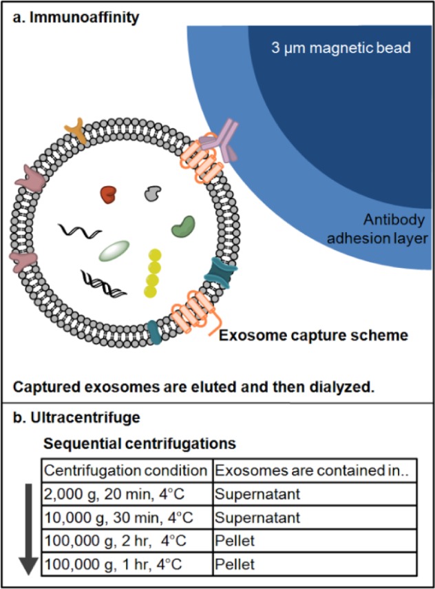
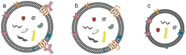
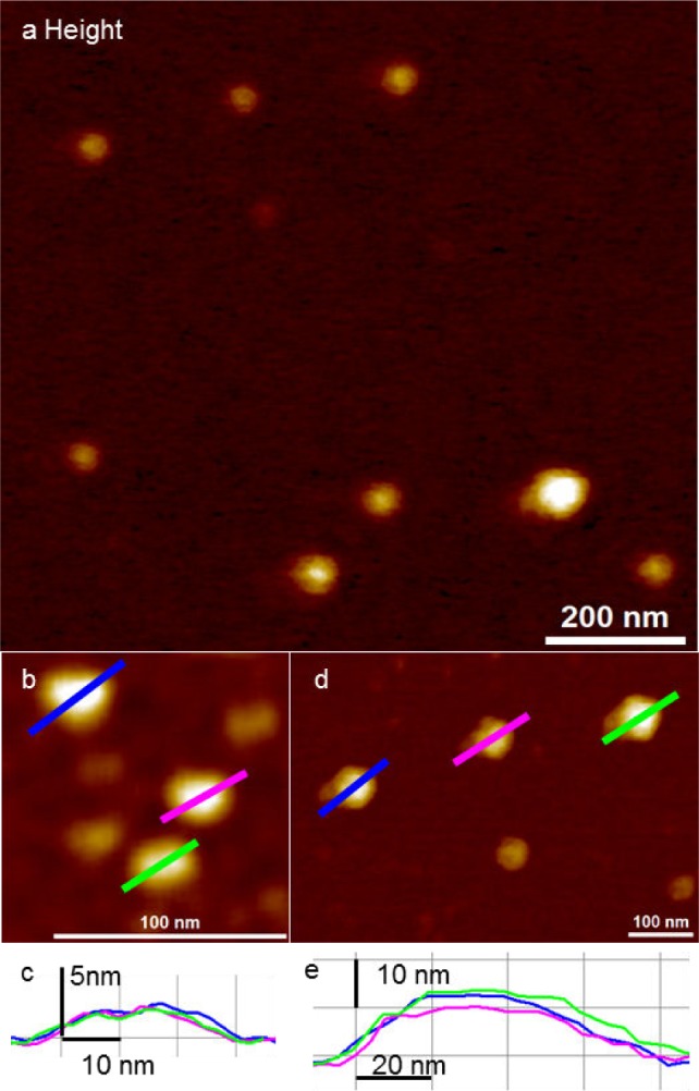
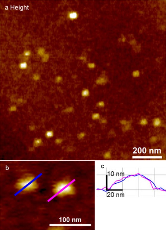
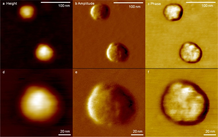
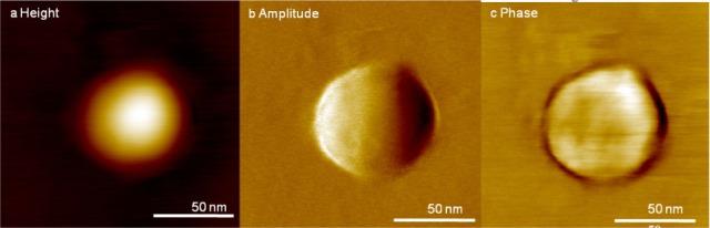
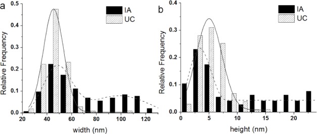
Exosomes are ∼100 nanometre diameter vesicles secreted by mammalian cells. These emerging disease biomarkers carry nucleic acids, proteins and lipids specific to the parental cells that secrete them. Exosomes are typically isolated in bulk by ultracentrifugation, filtration or immunoaffinity precipitation for downstream proteomic, genomic, or lipidomic analysis. However, the structural properties and heterogeneity of isolated exosomes at the single vesicle level are not well characterized due to their small size. In this paper, by using high-resolution atomic force microscope imaging, we show the nanoscale morphology and structural heterogeneity in exosomes derived from U87 cells. Quantitative assessment of single exosomes reveals nanoscale variations in morphology, surface roughness and counts isolated by ultracentrifugation (UC) and immunoaffinity (IA) purification. Both methods produce intact globular, 30-120 nm sized vesicles when imaged under fluid and in air. However, IA exosomes had higher surface roughness and bimodal size population compared to UC exosomes. The study highlights the differences in size and surface topography of exosomes purified from a single cell type using different isolation methods.

摘要

外泌体是由哺乳动物细胞分泌的直径约100纳米的囊泡。这些新兴的疾病生物标志物携带分泌它们的亲代细胞特有的核酸、蛋白质和脂质。外泌体通常通过超速离心、过滤或免疫亲和沉淀大量分离,用于下游的蛋白质组学、基因组学或脂质组学分析。然而,由于其尺寸小,在单囊泡水平上分离的外泌体的结构特性和异质性尚未得到很好的表征。在本文中,通过使用高分辨率原子力显微镜成像,我们展示了源自U87细胞的外泌体的纳米级形态和结构异质性。对单个外泌体的定量评估揭示了通过超速离心(UC)和免疫亲和(IA)纯化分离的外泌体在形态、表面粗糙度和数量上的纳米级变化。当在液体中和空气中成像时,这两种方法都产生完整的球形、大小为30 - 120纳米的囊泡。然而,与UC外泌体相比,IA外泌体具有更高的表面粗糙度和双峰大小分布。该研究突出了使用不同分离方法从单一细胞类型纯化的外泌体在大小和表面形貌上的差异。

https://cdn.ncbi.nlm.nih.gov/pmc/blobs/e797/5548320/58a7e53b01fb/10.5772_64148-fig7.jpg
https://cdn.ncbi.nlm.nih.gov/pmc/blobs/e797/5548320/1be9e0dcba4d/10.5772_64148-fig1.jpg
https://cdn.ncbi.nlm.nih.gov/pmc/blobs/e797/5548320/7764a3a04b5b/10.5772_64148-fig2.jpg
https://cdn.ncbi.nlm.nih.gov/pmc/blobs/e797/5548320/696382b57710/10.5772_64148-fig3.jpg
https://cdn.ncbi.nlm.nih.gov/pmc/blobs/e797/5548320/d3d568db7a2c/10.5772_64148-fig4.jpg
https://cdn.ncbi.nlm.nih.gov/pmc/blobs/e797/5548320/ae2d147b0c6a/10.5772_64148-fig5.jpg
https://cdn.ncbi.nlm.nih.gov/pmc/blobs/e797/5548320/7319a9b2108b/10.5772_64148-fig6.jpg
https://cdn.ncbi.nlm.nih.gov/pmc/blobs/e797/5548320/58a7e53b01fb/10.5772_64148-fig7.jpg

相似文献

1
The Role of Isolation Methods on a Nanoscale Surface Structure and its Effect on the Size of Exosomes.分离方法在纳米级表面结构上的作用及其对外泌体大小的影响。
J Circ Biomark. 2016 Jan 1;5:11. doi: 10.5772/64148. eCollection 2016 Jan-Dec.
2
Exosome Isolation by Ultracentrifugation and Precipitation and Techniques for Downstream Analyses.通过超速离心和沉淀法分离外泌体及下游分析技术
Curr Protoc Cell Biol. 2020 Sep;88(1):e110. doi: 10.1002/cpcb.110.
3
Comparison of exosomes purified via ultracentrifugation (UC) and Total Exosome Isolation (TEI) reagent from the serum of Marek's disease virus (MDV)-vaccinated and tumor-bearing chickens.比较通过超速离心(UC)和总外泌体分离(TEI)试剂从马立克氏病病毒(MDV)疫苗接种和肿瘤荷瘤鸡血清中纯化的外泌体。
J Virol Methods. 2019 Jan;263:1-9. doi: 10.1016/j.jviromet.2018.10.004. Epub 2018 Oct 11.
4
Evaluation of immune and chemical precipitation methods for plasma exosome isolation.评估用于血浆外泌体分离的免疫沉淀和化学沉淀方法。
PLoS One. 2020 Nov 24;15(11):e0242732. doi: 10.1371/journal.pone.0242732. eCollection 2020.
5
Integrated isolation and quantitative analysis of exosome shuttled proteins and nucleic acids using immunocapture approaches.使用免疫捕获方法对外泌体穿梭蛋白和核酸进行综合分离及定量分析。
Methods. 2015 Oct 1;87:46-58. doi: 10.1016/j.ymeth.2015.05.028. Epub 2015 Jun 2.
6
Plasma neuronal exosomes serve as biomarkers of cognitive impairment in HIV infection and Alzheimer's disease.血浆神经元外泌体可作为 HIV 感染和阿尔茨海默病认知障碍的生物标志物。
J Neurovirol. 2019 Oct;25(5):702-709. doi: 10.1007/s13365-018-0695-4. Epub 2019 Jan 4.
7
An improvised one-step sucrose cushion ultracentrifugation method for exosome isolation from culture supernatants of mesenchymal stem cells.一种改良的一步蔗糖垫超速离心法,用于从间充质干细胞培养上清液中分离外泌体。
Stem Cell Res Ther. 2018 Jul 4;9(1):180. doi: 10.1186/s13287-018-0923-0.
8
Quantitative nanostructural and single-molecule force spectroscopy biomolecular analysis of human-saliva-derived exosomes.人唾液衍生外泌体的定量纳米结构和单分子力谱生物分子分析。
Langmuir. 2011 Dec 6;27(23):14394-400. doi: 10.1021/la2038763. Epub 2011 Nov 9.
9
A protocol for exosome isolation and characterization: evaluation of ultracentrifugation, density-gradient separation, and immunoaffinity capture methods.外泌体分离与表征方案:超速离心、密度梯度分离和免疫亲和捕获方法的评估
Methods Mol Biol. 2015;1295:179-209. doi: 10.1007/978-1-4939-2550-6_15.
10
Identification and characterization of hADSC-derived exosome proteins from different isolation methods.不同分离方法来源的人脂肪间充质干细胞衍生外泌体蛋白的鉴定和特征分析。
J Cell Mol Med. 2021 Aug;25(15):7436-7450. doi: 10.1111/jcmm.16775. Epub 2021 Jul 8.

引用本文的文献

1
Exosomes: Methods for Isolation and Characterization in Biological Samples.外泌体:生物样本中分离和表征的方法。
Methods Mol Biol. 2024;2835:181-213. doi: 10.1007/978-1-0716-3995-5_17.
2
Therapeutic Potential of Bovine Milk-Derived Extracellular Vesicles.牛源细胞外囊泡的治疗潜力。
Int J Mol Sci. 2024 May 19;25(10):5543. doi: 10.3390/ijms25105543.
3
Combined Role of Interleukin-15 Stimulated Natural Killer Cell-Derived Extracellular Vesicles and Carboplatin in Osimertinib-Resistant H1975 Lung Cancer Cells with EGFR Mutations.

本文引用的文献

1
Proteomic analysis of exosomal cargo: the challenge of high purity vesicle isolation.外泌体货物的蛋白质组学分析:高纯度囊泡分离的挑战
Mol Biosyst. 2016 Apr 26;12(5):1407-19. doi: 10.1039/c6mb00082g.
2
Residual matrix from different separation techniques impacts exosome biological activity.来自不同分离技术的残留基质会影响外泌体的生物活性。
Sci Rep. 2016 Mar 24;6:23550. doi: 10.1038/srep23550.
3
Detection and isolation of circulating exosomes and microvesicles for cancer monitoring and diagnostics using micro-/nano-based devices.
白细胞介素-15刺激的自然杀伤细胞衍生的细胞外囊泡与卡铂在具有EGFR突变的奥希替尼耐药H1975肺癌细胞中的联合作用
Pharmaceutics. 2024 Jan 8;16(1):83. doi: 10.3390/pharmaceutics16010083.
4
Progress in Isolation and Molecular Profiling of Small Extracellular Vesicles via Bead-Assisted Platforms.基于微球辅助平台的小型细胞外囊泡的分离和分子特征分析的研究进展。
Biosensors (Basel). 2023 Jun 28;13(7):688. doi: 10.3390/bios13070688.
5
Endurance-dependent urinary extracellular vesicle signature: shape, metabolic miRNAs, and purine content distinguish triathletes from inactive people.耐力相关的尿细胞外囊泡特征:形态、代谢 miRNA 和嘌呤含量可区分运动员和非运动员。
Pflugers Arch. 2023 Jun;475(6):691-709. doi: 10.1007/s00424-023-02815-x. Epub 2023 May 8.
6
Assessment of anti-inflammatory bioactivity of extracellular vesicles is susceptible to error via media component contamination.评估细胞外囊泡的抗炎生物活性容易受到介质成分污染的影响而出现误差。
Cytotherapy. 2023 Apr;25(4):387-396. doi: 10.1016/j.jcyt.2022.12.002. Epub 2023 Jan 2.
7
Antenna-enhanced mid-infrared detection of extracellular vesicles derived from human cancer cell cultures.基于天线增强的人源癌细胞培养物衍生细胞外囊泡的中红外检测
J Nanobiotechnology. 2022 Dec 13;20(1):530. doi: 10.1186/s12951-022-01693-2.
8
Exosomes─Nature's Lipid Nanoparticles, a Rising Star in Drug Delivery and Diagnostics.外泌体——自然界的脂质纳米颗粒,药物传递和诊断领域的后起之秀。
ACS Nano. 2022 Nov 22;16(11):17802-17846. doi: 10.1021/acsnano.2c08774. Epub 2022 Nov 10.
9
Extracellular Vesicles Contribute to the Metabolism of Transthyretin Amyloid in Hereditary Transthyretin Amyloidosis.细胞外囊泡在遗传性转甲状腺素蛋白淀粉样变性中参与转甲状腺素蛋白淀粉样蛋白的代谢。
Front Mol Biosci. 2022 Mar 23;9:839917. doi: 10.3389/fmolb.2022.839917. eCollection 2022.
10
Overview of extracellular vesicle characterization techniques and introduction to combined reflectance and fluorescence confocal microscopy to distinguish extracellular vesicle subpopulations.细胞外囊泡表征技术概述以及结合反射和荧光共聚焦显微镜以区分细胞外囊泡亚群的介绍。
Neurophotonics. 2022 Apr;9(2):021903. doi: 10.1117/1.NPh.9.2.021903. Epub 2022 Apr 4.
使用基于微纳的装置进行循环外泌体和微囊泡的检测与分离,用于癌症监测和诊断。
Analyst. 2016 Jan 21;141(2):450-460. doi: 10.1039/c5an01610j.
4
Minimal experimental requirements for definition of extracellular vesicles and their functions: a position statement from the International Society for Extracellular Vesicles.最小实验要求定义细胞外囊泡及其功能:国际细胞外囊泡学会的立场声明。
J Extracell Vesicles. 2014 Dec 22;3:26913. doi: 10.3402/jev.v3.26913. eCollection 2014.
5
Magnetic bead-based isolation of exosomes.基于磁珠的外泌体分离
Methods Mol Biol. 2015;1218:465-81. doi: 10.1007/978-1-4939-1538-5_27.
6
Biogenesis, secretion, and intercellular interactions of exosomes and other extracellular vesicles.外泌体和其他细胞外囊泡的生物发生、分泌和细胞间相互作用。
Annu Rev Cell Dev Biol. 2014;30:255-89. doi: 10.1146/annurev-cellbio-101512-122326. Epub 2014 Aug 21.
7
Single-step isolation of extracellular vesicles by size-exclusion chromatography.通过大小排阻色谱法一步分离细胞外囊泡。
J Extracell Vesicles. 2014 Sep 8;3. doi: 10.3402/jev.v3.23430. eCollection 2014.
8
Tetraspanins in extracellular vesicle formation and function.四跨膜蛋白在细胞外囊泡形成和功能中的作用。
Front Immunol. 2014 Sep 16;5:442. doi: 10.3389/fimmu.2014.00442. eCollection 2014.
9
Particle size distribution of exosomes and microvesicles determined by transmission electron microscopy, flow cytometry, nanoparticle tracking analysis, and resistive pulse sensing.通过透射电子显微镜、流式细胞术、纳米颗粒跟踪分析和电阻脉冲感应测定外泌体和微泡的粒径分布。
J Thromb Haemost. 2014 Jul;12(7):1182-92. doi: 10.1111/jth.12602. Epub 2014 Jun 19.
10
Nanofilaments on glioblastoma exosomes revealed by peak force microscopy.通过峰值力显微镜揭示神经胶母细胞瘤外泌体上的纳米丝。
J R Soc Interface. 2014 Jan 8;11(92):20131150. doi: 10.1098/rsif.2013.1150. Print 2014 Mar 6.