Department of Biology, University of Central Arkansas Conway, Conway, Arkansas.
Department of Pharmacology and Toxicology, College of Medicine, University of Arkansas for Medical Sciences, Little Rock, Arkansas.
Pharmacol Res Perspect. 2017 Oct;5(5). doi: 10.1002/prp2.358.
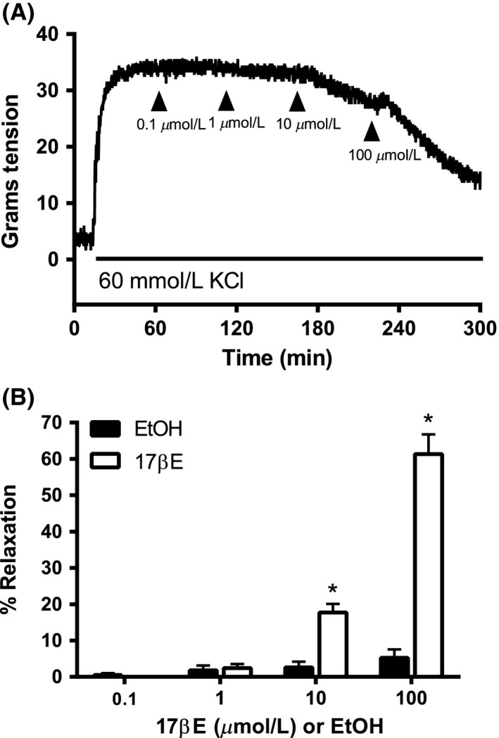
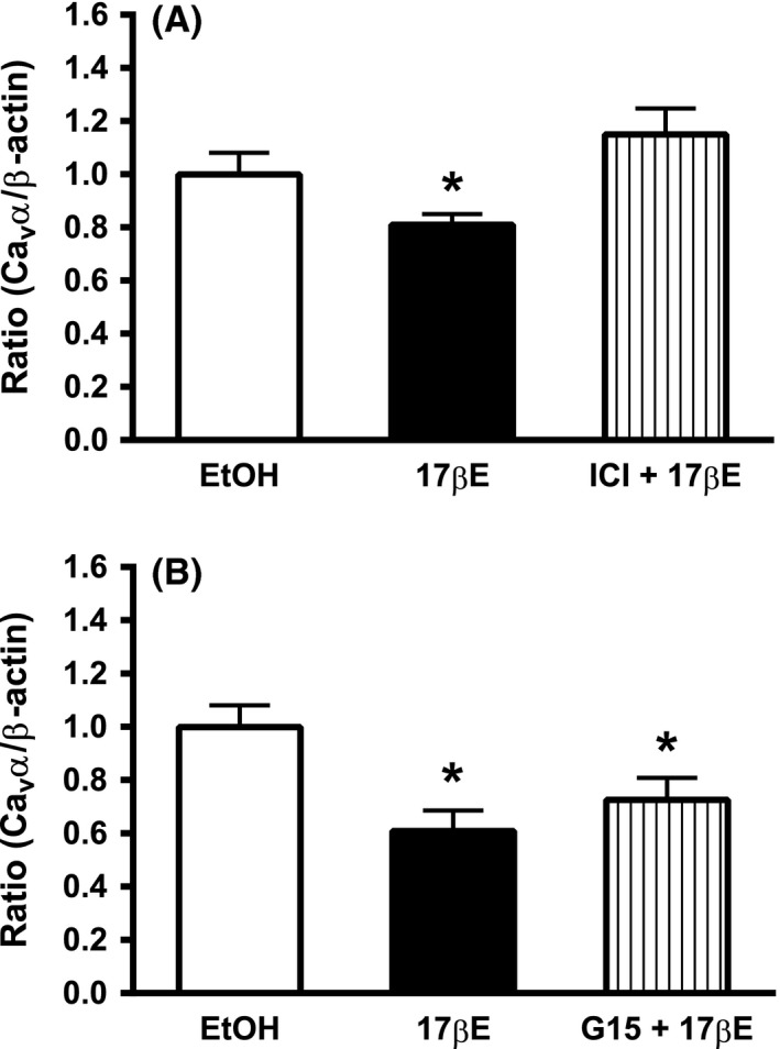
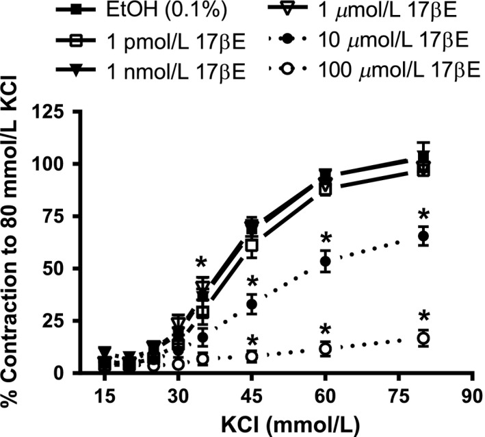
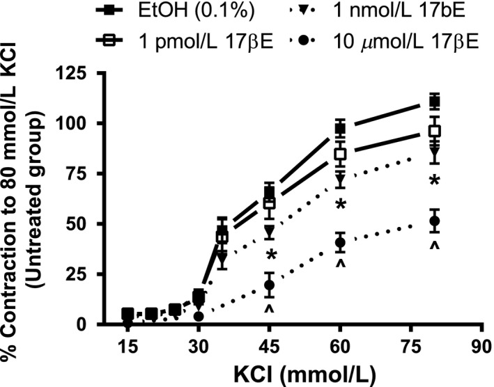
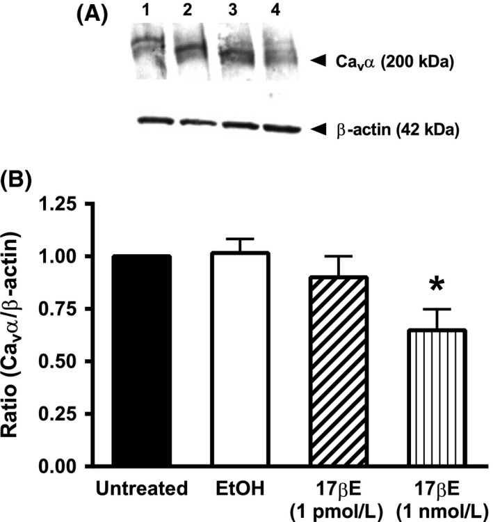
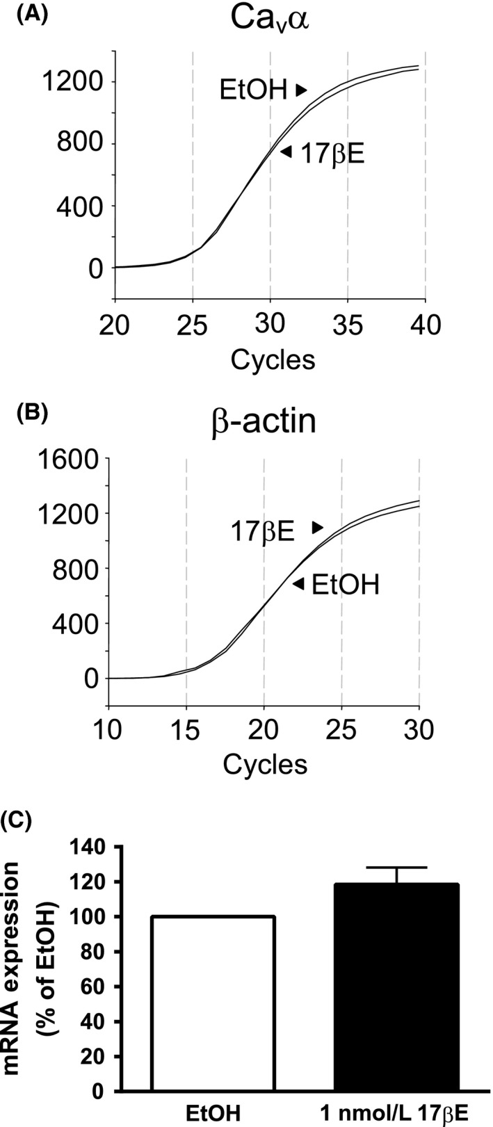
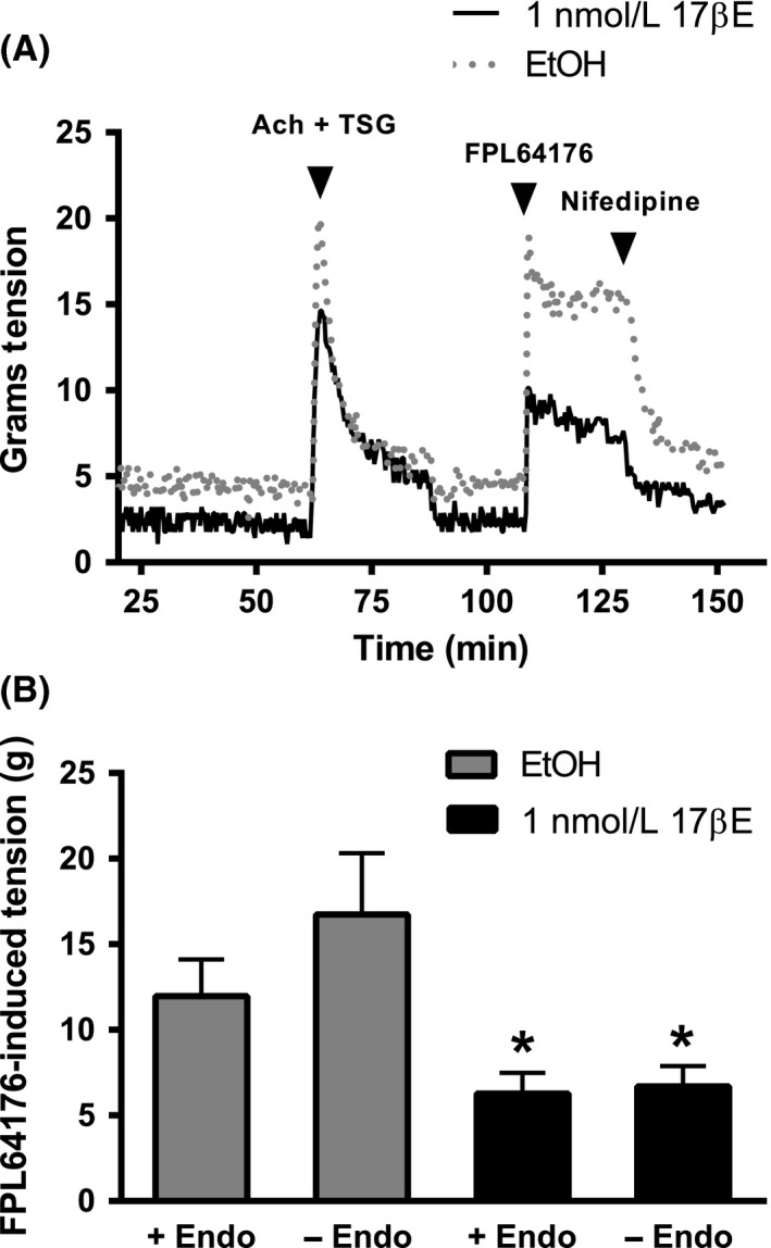
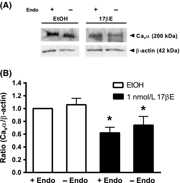
One mechanism by which the female sex may protect against elevated coronary vascular tone is inhibition of Ca entry into arterial smooth muscle cells (ASMCs). In vitro findings confirm that high estrogen concentrations directly inhibit voltage-dependent Ca 1.2 channels in coronary ASMCs. For this study, we hypothesized that the nonacute, in vitro exposure of coronary arteries to a low concentration of 17β-estradiol (17βE) reduces the expression of Ca 1.2 channel proteins in coronary ASMCs. Segments of the right coronary artery obtained from sexually mature female pigs were mounted for isometric tension recording. As expected, our results indicate that high concentrations (≥10 μmol/L) of 17βE acutely attenuated Ca -dependent contractions to depolarizing KCl stimuli. Interestingly, culturing coronary arteries for 24 h in a 10,000-fold lower concentration (1 nmol/L) of 17βE also attenuated KCl-induced contractions and reduced the contractile response to the Ca 1.2 agonist, FPL64176, by 50%. Western blots revealed that 1 nmol/L 17βE decreased protein expression of the pore-forming α subunit (Ca α) of the Ca 1.2 channel by 35%; this response did not depend on an intact endothelium. The 17βE-induced loss of Ca α protein in coronary arteries was prevented by the estrogen ERα/ERβ antagonist, ICI 182,780, whereas the GPER antagonist, G15, did not prevent it. There was no effect of 1 nmol/L 17βE on Ca α transcript expression. We conclude that 17βE reduces Ca 1.2 channel abundance in isolated coronary arteries by a posttranscriptional process. This unrecognized effect of estrogen may confer physiological protection against the development of abnormal Ca -dependent coronary vascular tone.
女性可能通过抑制动脉平滑肌细胞(ASMC)内钙离子内流来保护冠状血管免受升高的血管张力。体外研究结果证实,高浓度雌激素可直接抑制冠状动脉 ASMC 中的电压依赖性钙通道。在这项研究中,我们假设,在体外,非急性、低浓度的 17β-雌二醇(17βE)暴露于冠状动脉会降低冠状动脉 ASMC 中钙通道 1.2 型蛋白的表达。从性成熟雌性猪获得的右冠状动脉段被安装在等长张力记录器上。正如预期的那样,我们的结果表明,高浓度(≥10μmol/L)的 17βE 可急性减弱去极化 KCl 刺激引起的 Ca2+依赖性收缩。有趣的是,在低浓度(1nmol/L)的 17βE 中培养冠状动脉 24 小时也可减弱 KCl 引起的收缩,并使 Ca1.2 激动剂 FPL64176 的收缩反应降低 50%。Western blot 显示,1nmol/L 的 17βE 使钙通道的孔形成α亚基(Caα)的蛋白表达减少 35%;这种反应不依赖于完整的内皮。雌激素 ERα/ERβ拮抗剂 ICI 182,780 可阻止 17βE 诱导的冠状动脉 Caα蛋白丢失,而 GPER 拮抗剂 G15 则不能阻止。1nmol/L 的 17βE 对 Caα转录物的表达没有影响。我们的结论是,17βE 通过转录后过程减少了分离的冠状动脉中钙通道 1.2 的丰度。这种雌激素的未被认识的作用可能为异常 Ca2+依赖性冠状血管张力的发展提供了生理性保护。