• 文献检索
  • 文档翻译
  • 深度研究
  • 学术资讯
  • Suppr Zotero 插件Zotero 插件
  • 邀请有礼
  • 套餐&价格
  • 历史记录
应用&插件
Suppr Zotero 插件Zotero 插件浏览器插件Mac 客户端Windows 客户端微信小程序
定价
高级版会员购买积分包购买API积分包
服务
文献检索文档翻译深度研究API 文档MCP 服务
关于我们
关于 Suppr公司介绍联系我们用户协议隐私条款
关注我们

Suppr 超能文献

核心技术专利:CN118964589B侵权必究
粤ICP备2023148730 号-1Suppr @ 2026

文献检索

告别复杂PubMed语法,用中文像聊天一样搜索,搜遍4000万医学文献。AI智能推荐,让科研检索更轻松。

立即免费搜索

文件翻译

保留排版,准确专业,支持PDF/Word/PPT等文件格式,支持 12+语言互译。

免费翻译文档

深度研究

AI帮你快速写综述,25分钟生成高质量综述,智能提取关键信息,辅助科研写作。

立即免费体验

成年小鼠中骨形态发生蛋白2诱导的中指骨再生的骨膜需求和时间动态

The periosteal requirement and temporal dynamics of BMP2-induced middle phalanx regeneration in the adult mouse.

作者信息

Dawson Lindsay A, Yu Ling, Yan Mingquan, Marrero Luis, Schanes Paula P, Dolan Connor, Pela Maegan, Petersen Britta, Han Manjong, Muneoka Ken

机构信息

Veterinary Physiology and Pharmacology Texas A&M University 4466 TAMU College Station TX USA.

Cell and Molecular Biology Tulane University 2000 Percival Stern Hall, 6400 Freret St New Orleans LA USA.

出版信息

Regeneration (Oxf). 2017 Aug 20;4(3):140-150. doi: 10.1002/reg2.81. eCollection 2017 Jun.

DOI:10.1002/reg2.81
PMID:28975034
原文链接:https://pmc.ncbi.nlm.nih.gov/articles/PMC5617898/
Abstract

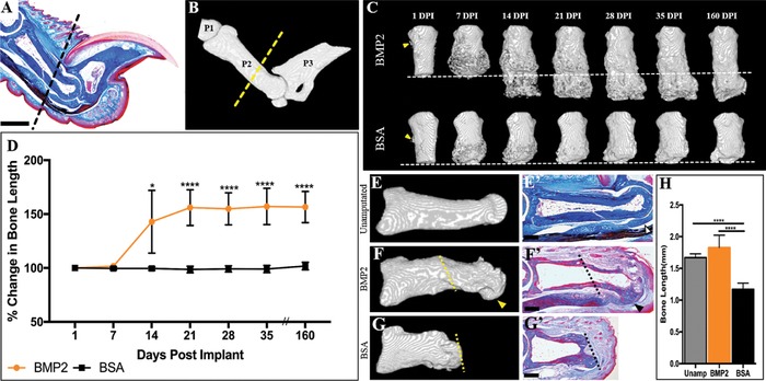
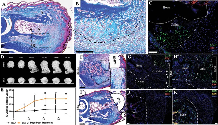
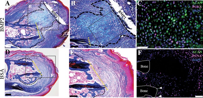
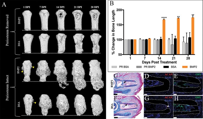
Regeneration of mammalian limbs is restricted to amputation of the distal digit tip, the terminal phalanx (P3). The adjacent skeletal element, the middle phalanx (P2), has emerged as a model system to investigate regenerative failure and as a site to test approaches aimed at enhancing regeneration. We report that exogenous application of bone morphogenetic protein 2 (BMP2) stimulates the formation of a transient cartilaginous callus distal to the amputation plane that mediates the regeneration of the amputated P2 bone. BMP2 initiates a significant regeneration response during the periosteal-derived cartilaginous healing phase of P2 bone repair, yet fails to induce regeneration in the absence of periosteal tissue, or after boney callus formation. We provide evidence that a temporal component exists in the induced regeneration of P2 that we define as the "regeneration window." In this window, cells are transiently responsive to BMP2 after the amputation injury. Simple re-injury of the healed P2 stump acts to reinitiate endogenous bone repair, complete with periosteal chondrogenesis, thus reopening the "regeneration window" and thereby recreating a regeneration-permissive environment that is responsive to exogenous BMP2 treatment.

摘要

哺乳动物肢体的再生仅限于远端指尖(即末节指骨,P3)的截肢情况。相邻的骨骼元件,即中节指骨(P2),已成为研究再生失败的模型系统,也是测试旨在增强再生方法的部位。我们报告称,外源性应用骨形态发生蛋白2(BMP2)可刺激在截肢平面远端形成一个短暂的软骨痂,该软骨痂介导了截肢的P2骨的再生。BMP2在P2骨修复的骨膜来源的软骨愈合阶段引发显著的再生反应,但在没有骨膜组织的情况下或在骨痂形成后无法诱导再生。我们提供证据表明,在P2的诱导再生中存在一个时间成分,我们将其定义为“再生窗口”。在此窗口内,截肢损伤后细胞对BMP2产生短暂反应。对愈合的P2残端进行简单的再次损伤可重新启动内源性骨修复,包括骨膜软骨形成,从而重新打开“再生窗口”,进而重新创造一个对外源性BMP2治疗有反应的再生允许环境。

https://cdn.ncbi.nlm.nih.gov/pmc/blobs/0c49/5617898/11b1de162b30/REG2-4-140-g005.jpg
https://cdn.ncbi.nlm.nih.gov/pmc/blobs/0c49/5617898/8b554abc945c/REG2-4-140-g001.jpg
https://cdn.ncbi.nlm.nih.gov/pmc/blobs/0c49/5617898/52fc8b081266/REG2-4-140-g002.jpg
https://cdn.ncbi.nlm.nih.gov/pmc/blobs/0c49/5617898/abfecfb040aa/REG2-4-140-g003.jpg
https://cdn.ncbi.nlm.nih.gov/pmc/blobs/0c49/5617898/8b2664b33829/REG2-4-140-g004.jpg
https://cdn.ncbi.nlm.nih.gov/pmc/blobs/0c49/5617898/11b1de162b30/REG2-4-140-g005.jpg

相似文献

1
The periosteal requirement and temporal dynamics of BMP2-induced middle phalanx regeneration in the adult mouse.成年小鼠中骨形态发生蛋白2诱导的中指骨再生的骨膜需求和时间动态
Regeneration (Oxf). 2017 Aug 20;4(3):140-150. doi: 10.1002/reg2.81. eCollection 2017 Jun.
2
BMP2 induces segment-specific skeletal regeneration from digit and limb amputations by establishing a new endochondral ossification center.BMP2 通过建立新的软骨内骨化中心,诱导来自手指和肢体截肢的节段特异性骨骼再生。
Dev Biol. 2012 Dec 15;372(2):263-73. doi: 10.1016/j.ydbio.2012.09.021. Epub 2012 Oct 3.
3
Analogous cellular contribution and healing mechanisms following digit amputation and phalangeal fracture in mice.小鼠断指与指骨骨折后类似的细胞贡献和愈合机制。
Regeneration (Oxf). 2016 Mar 9;3(1):39-51. doi: 10.1002/reg2.51. eCollection 2016 Feb.
4
Proximal digit tip amputation initiates simultaneous blastema and transient fibrosis formation and results in partial regeneration.近端指端截断会同时引发芽基和短暂纤维化的形成,从而导致部分再生。
Wound Repair Regen. 2021 Jan;29(1):196-205. doi: 10.1111/wrr.12856. Epub 2020 Aug 21.
5
Blastema formation and periosteal ossification in the regenerating adult mouse digit.成年小鼠再生指中芽基的形成和骨膜骨化
Wound Repair Regen. 2018 May;26(3):263-273. doi: 10.1111/wrr.12666. Epub 2018 Oct 16.
6
Bone morphogenetic protein 2 stimulates endochondral ossification by regulating periosteal cell fate during bone repair.骨形态发生蛋白 2 通过调节骨修复过程中骨膜细胞的命运来刺激软骨内骨化。
Bone. 2010 Jul;47(1):65-73. doi: 10.1016/j.bone.2010.03.012. Epub 2010 Mar 27.
7
Periosteal BMP2 activity drives bone graft healing.骨膜 BMP2 活性驱动骨移植物愈合。
Bone. 2012 Oct;51(4):800-9. doi: 10.1016/j.bone.2012.07.017. Epub 2012 Jul 28.
8
Connective tissue fibroblast properties are position-dependent during mouse digit tip regeneration.结缔组织成纤维细胞在小鼠指尖再生过程中的位置依赖性。
PLoS One. 2013;8(1):e54764. doi: 10.1371/journal.pone.0054764. Epub 2013 Jan 18.
9
BMP signaling induces digit regeneration in neonatal mice.BMP 信号诱导新生小鼠的趾再生。
Development. 2010 Feb;137(4):551-9. doi: 10.1242/dev.042424.
10
Adult Mouse Digit Amputation and Regeneration: A Simple Model to Investigate Mammalian Blastema Formation and Intramembranous Ossification.成年小鼠指趾截肢与再生:用于研究哺乳动物芽基形成和膜内成骨的简单模型
J Vis Exp. 2019 Jul 12(149). doi: 10.3791/59749.

引用本文的文献

1
Digit regeneration is expedited in LG/J healer mice compared to SM/J non-healer mice.与SM/J非愈合型小鼠相比,LG/J愈合型小鼠的趾再生速度更快。
NPJ Regen Med. 2025 Feb 15;10(1):11. doi: 10.1038/s41536-025-00399-x.
2
The guanine nucleotide exchange factor DOCK5 negatively regulates osteoblast differentiation and BMP2-induced bone regeneration via the MKK3/6 and p38 signaling pathways.鸟嘌呤核苷酸交换因子DOCK5通过MKK3/6和p38信号通路负向调节成骨细胞分化和BMP2诱导的骨再生。
Exp Mol Med. 2025 Feb;57(1):86-103. doi: 10.1038/s12276-024-01372-2. Epub 2025 Jan 1.
3
Male Down syndrome Ts65Dn mice have impaired bone regeneration.

本文引用的文献

1
Macrophages are required to coordinate mouse digit tip regeneration.巨噬细胞是协调小鼠指尖再生所必需的。
Development. 2017 Nov 1;144(21):3907-3916. doi: 10.1242/dev.150086. Epub 2017 Sep 21.
2
The biology of bone lengthening.骨延长生物学
J Child Orthop. 2016 Dec;10(6):487-492. doi: 10.1007/s11832-016-0780-2. Epub 2016 Nov 12.
3
Analogous cellular contribution and healing mechanisms following digit amputation and phalangeal fracture in mice.小鼠断指与指骨骨折后类似的细胞贡献和愈合机制。
雄性唐氏综合征 Ts65Dn 小鼠的骨再生受损。
Bone. 2025 Mar;192:117374. doi: 10.1016/j.bone.2024.117374. Epub 2024 Dec 13.
4
Deer antler renewal gives insights into mammalian epimorphic regeneration.鹿茸再生为了解哺乳动物的表皮形态再生提供了线索。
Cell Regen. 2023 Jul 25;12(1):26. doi: 10.1186/s13619-023-00169-4.
5
Toeing the line between regeneration and fibrosis.在再生与纤维化之间保持平衡。
Front Cell Dev Biol. 2023 Jun 1;11:1217185. doi: 10.3389/fcell.2023.1217185. eCollection 2023.
6
Single-cell mapping of regenerative and fibrotic healing responses after musculoskeletal injury.骨骼肌损伤后再生和纤维化修复反应的单细胞图谱绘制。
Stem Cell Reports. 2022 Oct 11;17(10):2334-2348. doi: 10.1016/j.stemcr.2022.08.011. Epub 2022 Sep 22.
7
Evaluating differences in Young's Modulus of regenerated and uninjured mouse digit bone through microCT density-based calculation and nanoindentation testing.通过 microCT 密度计算和纳米压痕测试评估再生和未受伤的小鼠趾骨杨氏模量的差异。
J Biomech. 2022 Oct;143:111271. doi: 10.1016/j.jbiomech.2022.111271. Epub 2022 Aug 27.
8
To regenerate or not to regenerate: Vertebrate model organisms of regeneration-competency and -incompetency.是否再生:具有再生能力和无再生能力的脊椎动物模式生物。
Wound Repair Regen. 2022 Nov;30(6):623-635. doi: 10.1111/wrr.13000. Epub 2022 Feb 22.
9
Hyaline cartilage differentiation of fibroblasts in regeneration and regenerative medicine.纤维母细胞在再生和再生医学中的透明软骨分化。
Development. 2022 Jan 15;149(2). doi: 10.1242/dev.200249. Epub 2022 Jan 28.
10
Mouse Digit Tip Regeneration Is Mechanical Load Dependent.鼠标指尖再生依赖于机械负荷。
J Bone Miner Res. 2022 Feb;37(2):312-322. doi: 10.1002/jbmr.4470. Epub 2021 Dec 7.
Regeneration (Oxf). 2016 Mar 9;3(1):39-51. doi: 10.1002/reg2.51. eCollection 2016 Feb.
4
Skeletal callus formation is a nerve-independent regenerative response to limb amputation in mice and Xenopus.骨骼骨痂形成是小鼠和非洲爪蟾对肢体截肢的一种不依赖神经的再生反应。
Regeneration (Oxf). 2015 Aug 26;2(4):202-16. doi: 10.1002/reg2.39. eCollection 2015 Aug.
5
Epidermal closure regulates histolysis during mammalian (Mus) digit regeneration.表皮闭合调节哺乳动物(小家鼠)趾再生过程中的组织溶解。
Regeneration (Oxf). 2015 Jun 9;2(3):106-19. doi: 10.1002/reg2.34. eCollection 2015 Jun.
6
The mammalian blastema: regeneration at our fingertips.哺乳动物再生芽基:触手可及的再生
Regeneration (Oxf). 2015 Jun 9;2(3):93-105. doi: 10.1002/reg2.36. eCollection 2015 Jun.
7
The Molecular and Cellular Choreography of Appendage Regeneration.附肢再生的分子和细胞舞蹈。
Cell. 2016 Jun 16;165(7):1598-1608. doi: 10.1016/j.cell.2016.05.038.
8
A review on carrier systems for bone morphogenetic protein-2.骨形态发生蛋白-2载体系统综述。
J Biomed Mater Res B Appl Biomater. 2017 May;105(4):904-925. doi: 10.1002/jbm.b.33599. Epub 2016 Jan 5.
9
Review of the cellular and biological principles of distraction osteogenesis: An in vivo bioreactor tissue engineering model.牵张成骨的细胞与生物学原理综述:一种体内生物反应器组织工程模型
J Plast Reconstr Aesthet Surg. 2016 Feb;69(2):e19-26. doi: 10.1016/j.bjps.2015.11.003. Epub 2015 Nov 25.
10
Hyperbaric Oxygen Promotes Proximal Bone Regeneration and Organized Collagen Composition during Digit Regeneration.高压氧促进指(趾)再生过程中的近端骨再生和有序胶原组成。
PLoS One. 2015 Oct 9;10(10):e0140156. doi: 10.1371/journal.pone.0140156. eCollection 2015.