Li Wei-Wei, Wang Hai-Yue, Nie Xi, Liu Ya-Bin, Han Mei, Li Bing-Hui
Department of Biochemistry and Molecular Biology, College of Basic Medicine, Key Laboratory of Medical Biotechnology of Hebei Province, Shijiazhuang 050017, P. R. China.
Department of Surgery, Fourth Affiliated Hospital, Hebei Medical University, Shijiazhuang 050017, P. R. China.
Oncotarget. 2017 Jun 27;8(37):62049-62056. doi: 10.18632/oncotarget.18893. eCollection 2017 Sep 22.
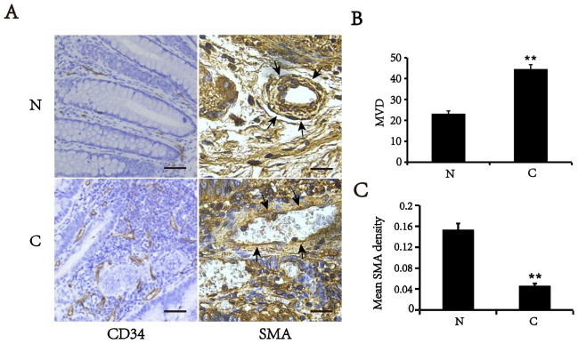
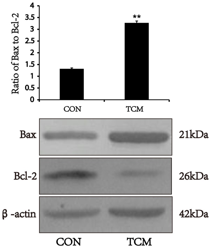
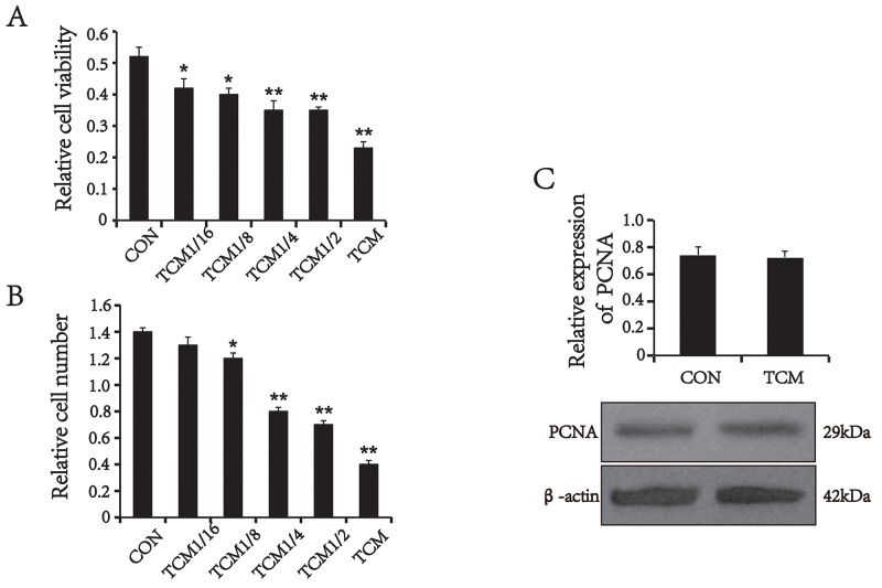
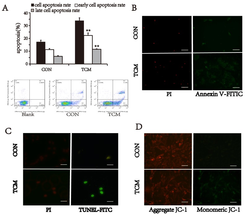
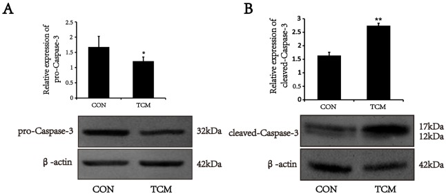
Tumor vessels often lack the smooth muscle layer, and the instability is conducive to tumor invasion and metastasis. The effect of tumor microenvironment on vascular smooth muscle cells needs to be explored. In the present study, we examined the density of the tumor vessels in human colorectal cancer tissues, and used the tumor conditioned medium of human colorectal cancer HT29 cells to mimic the tumor microenvironment. We showed that the vessel density in colorectal cancer tissues increased, which displayed a decreased expression of smooth muscle α-actin, a specific marker of vascular smooth muscle cells and an attenuated or a discontinuous layer of vascular smooth muscle cells compared with the matched normal tissues. We also showed that the tumor conditioned medium decreased the cell viability, and induced the apoptosis in vascular smooth muscle cells in a concentration-dependent manner. The expression of pro-Caspase-3 was down-regulated, accompanied by increasing of cleaved-Caspase-3 in the cells treated with the tumor conditioned medium, suggesting that Caspase-3 was activated. Moreover, the expression of Bax was increased, and the ratio of Bcl-2/Bax was decreased under the same conditions. Furthermore, the treatment with the tumor conditioned medium resulted in loss of mitochondrial membrane potential in vascular smooth muscle cells. These findings suggest that HT29 cells induce apoptosis of vascular smooth muscle cells in an exocrine manner, associated with activating caspase-3 via mitochondrial apoptotic pathway. This may be one of the mechanisms underlying tumor vascular structural abnormalities.
肿瘤血管通常缺乏平滑肌层,这种不稳定性有利于肿瘤的侵袭和转移。肿瘤微环境对血管平滑肌细胞的影响有待探索。在本研究中,我们检测了人结直肠癌组织中肿瘤血管的密度,并用人结直肠癌HT29细胞的肿瘤条件培养基模拟肿瘤微环境。我们发现,结直肠癌组织中的血管密度增加,与配对的正常组织相比,血管平滑肌细胞的特异性标志物平滑肌α-肌动蛋白表达降低,血管平滑肌细胞层变薄或不连续。我们还发现,肿瘤条件培养基以浓度依赖的方式降低了血管平滑肌细胞的活力并诱导其凋亡。在用肿瘤条件培养基处理的细胞中,前半胱天冬酶-3的表达下调,同时裂解的半胱天冬酶-3增加,这表明半胱天冬酶-3被激活。此外,在相同条件下,Bax的表达增加,Bcl-2/Bax的比值降低。此外,用肿瘤条件培养基处理导致血管平滑肌细胞线粒体膜电位丧失。这些发现表明,HT29细胞以外分泌方式诱导血管平滑肌细胞凋亡,这与通过线粒体凋亡途径激活半胱天冬酶-3有关。这可能是肿瘤血管结构异常的潜在机制之一。