Bone and Signaling Laboratory, Space BioSciences Division, NASA Ames Research Center, Mail-Stop 236-7, Moffett Field, CA 94035, USA.
Wyle Laboratories, Mail-Stop 236-7, Moffett Field, CA 94035, USA.
Int J Mol Sci. 2017 Oct 10;18(10):2117. doi: 10.3390/ijms18102117.
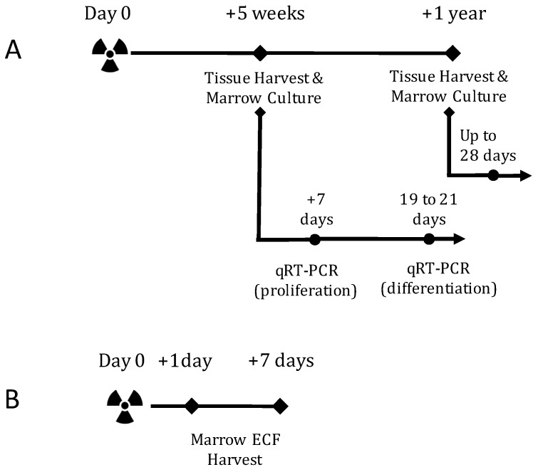
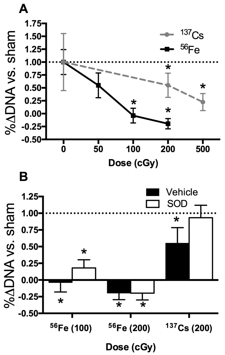
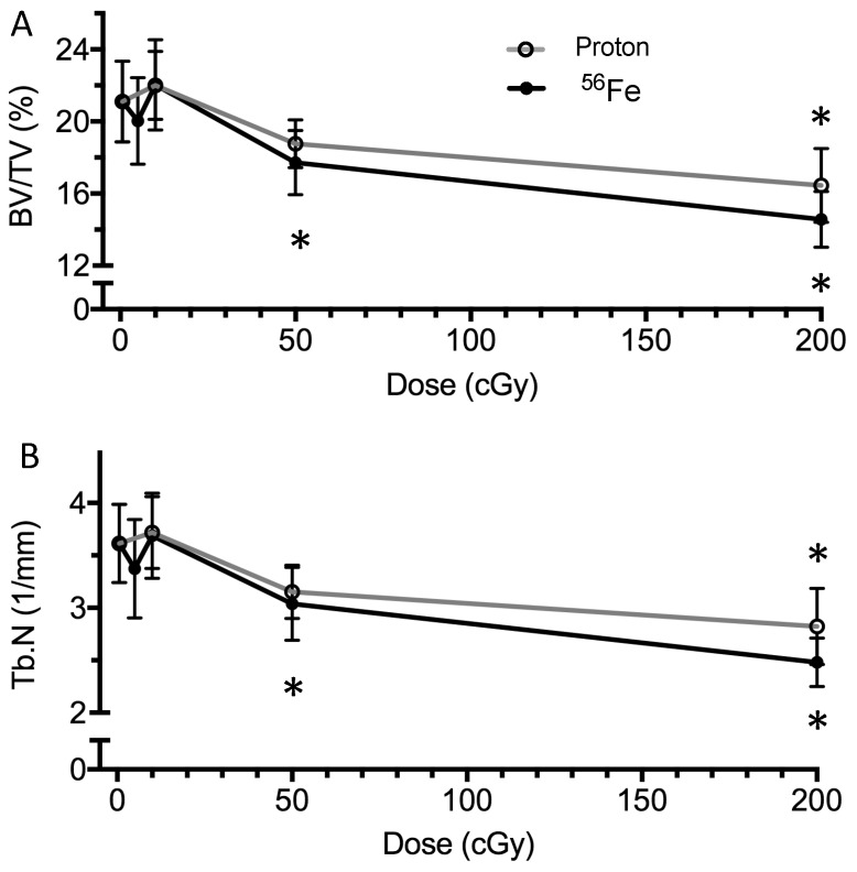
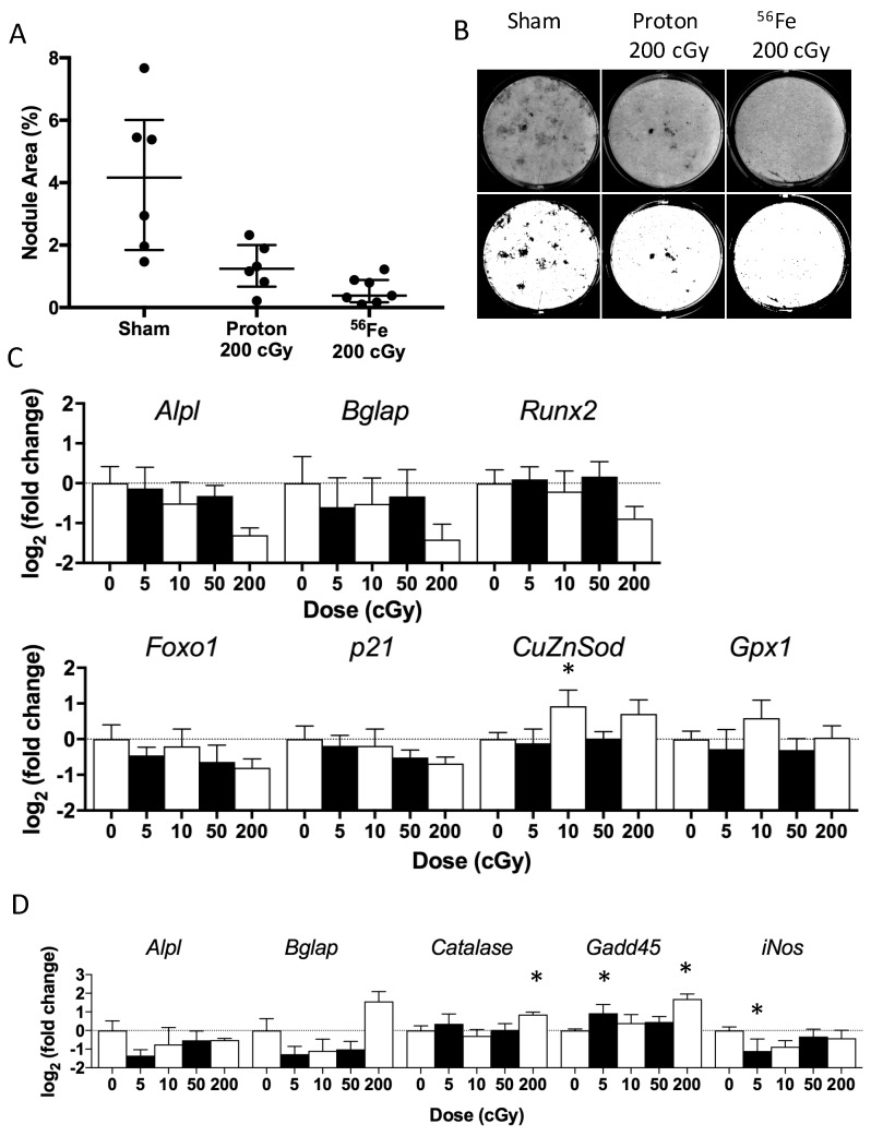
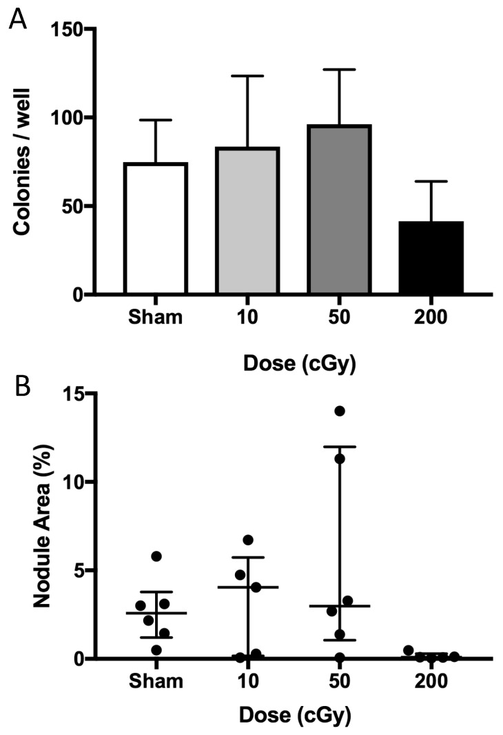
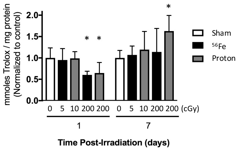
Space radiation may pose a risk to skeletal health during subsequent aging. Irradiation acutely stimulates bone remodeling in mice, although the long-term influence of space radiation on bone-forming potential (osteoblastogenesis) and possible adaptive mechanisms are not well understood. We hypothesized that ionizing radiation impairs osteoblastogenesis in an ion-type specific manner, with low doses capable of modulating expression of redox-related genes. 16-weeks old, male, C57BL6/J mice were exposed to low linear-energy-transfer (LET) protons (150 MeV/n) or high-LET Fe ions (600 MeV/n) using either low (5 or 10 cGy) or high (50 or 200 cGy) doses at NASA's Space Radiation Lab. Five weeks or one year after irradiation, tissues were harvested and analyzed by microcomputed tomography for cancellous microarchitecture and cortical geometry. Marrow-derived, adherent cells were grown under osteoblastogenic culture conditions. Cell lysates were analyzed by RT-PCR during the proliferative or mineralizing phase of growth, and differentiation was analyzed by imaging mineralized nodules. As expected, a high dose (200 cGy), but not lower doses, of either Fe or protons caused a loss of cancellous bone volume/total volume. Marrow cells produced mineralized nodules ex vivo regardless of radiation type or dose; Fe (200 cGy) inhibited osteoblastogenesis by more than 90% (5 weeks and 1 year post-IR). After 5 weeks, irradiation (protons or Fe) caused few changes in gene expression levels during osteoblastogenesis, although a high dose Fe (200 cGy) increased and . The addition of exogenous superoxide dismutase (SOD) protected marrow-derived osteoprogenitors from the damaging effects of exposure to low-LET (Cs γ) when irradiated in vitro, but had limited protective effects on high-LET Fe-exposed cells. In sum, either protons or Fe at a relatively high dose (200 cGy) caused persistent bone loss, whereas only high-LET Fe increased redox-related gene expression, albeit to a limited extent, and inhibited osteoblastogenesis. Doses below 50 cGy did not elicit widespread responses in any parameter measured. We conclude that high-LET irradiation at 200 cGy impaired osteoblastogenesis and regulated steady-state gene expression of select redox-related genes during osteoblastogenesis, which may contribute to persistent bone loss.
空间辐射可能会对后续衰老过程中的骨骼健康造成风险。辐照会在小鼠体内急性刺激骨重塑,尽管目前还不清楚空间辐射对成骨潜能(成骨细胞发生)和可能的适应机制的长期影响。我们假设,电离辐射会以特定的离子类型方式损害成骨细胞发生,低剂量能够调节氧化还原相关基因的表达。16 周龄的雄性 C57BL6/J 小鼠使用 NASA 空间辐射实验室中的低线性能量转移(LET)质子(150 MeV/n)或高 LET Fe 离子(600 MeV/n),接受 5 或 10 cGy 或 50 或 200 cGy 的低或高剂量照射。照射后 5 周或 1 年,采集组织,通过微计算机断层扫描分析松质骨微结构和皮质几何形状。骨髓来源的贴壁细胞在成骨细胞培养条件下生长。在细胞增殖或矿化阶段,通过 RT-PCR 分析细胞裂解物,通过成像矿化结节分析分化。正如预期的那样,高剂量(200 cGy),但不是低剂量的 Fe 或质子都会导致松质骨体积/总体积丢失。无论辐射类型或剂量如何,骨髓细胞都会在体外产生矿化结节;Fe(200 cGy)抑制成骨细胞发生超过 90%(辐照后 5 周和 1 年)。照射后 5 周,辐照(质子或 Fe)在成骨细胞发生过程中引起的基因表达水平变化很少,尽管高剂量 Fe(200 cGy)增加了 和 。当体外辐照时,添加外源性超氧化物歧化酶(SOD)可以保护骨髓来源的成骨祖细胞免受低 LET(Cs γ)的损伤,但对高 LET Fe 暴露细胞的保护作用有限。总之,相对高剂量(200 cGy)的质子或 Fe 会导致持续的骨丢失,而只有高 LET Fe 会增加氧化还原相关基因的表达,尽管程度有限,但会抑制成骨细胞发生。低于 50 cGy 的剂量不会引起任何测量参数的广泛反应。我们得出结论,200 cGy 的高 LET 辐照会损害成骨细胞发生,并调节成骨细胞发生过程中成骨相关基因的稳态基因表达,这可能导致持续的骨丢失。